
Effects of sub-chronic, in vivo administration of sigma non-opioid intracellular receptor 1 ligands on platelet and aortic arachidonate cascade in rats
By Sándor Váczi, Lilla Barna, Krisztián Laczi, Ferenc Tömösi, Gábor Rákhely, Botond Penke, Lívia Fülöp, Ferenc Bogár, Tamás Janáky, Mária A. Deli, and Zsófia Mezei
Excerpt from the article published in European Journal of Pharmacology, 2022, 174983 Doi: https://doi.org/10.1016/j.ejphar.2022.174983.
Editor’s Highlights
- Only a fraction of all cardiovascular diseases is of cardiac origin, and macro-and microvascular dysfunction and/or platelet activation are responsible for the development and exacerbation of many diseases.
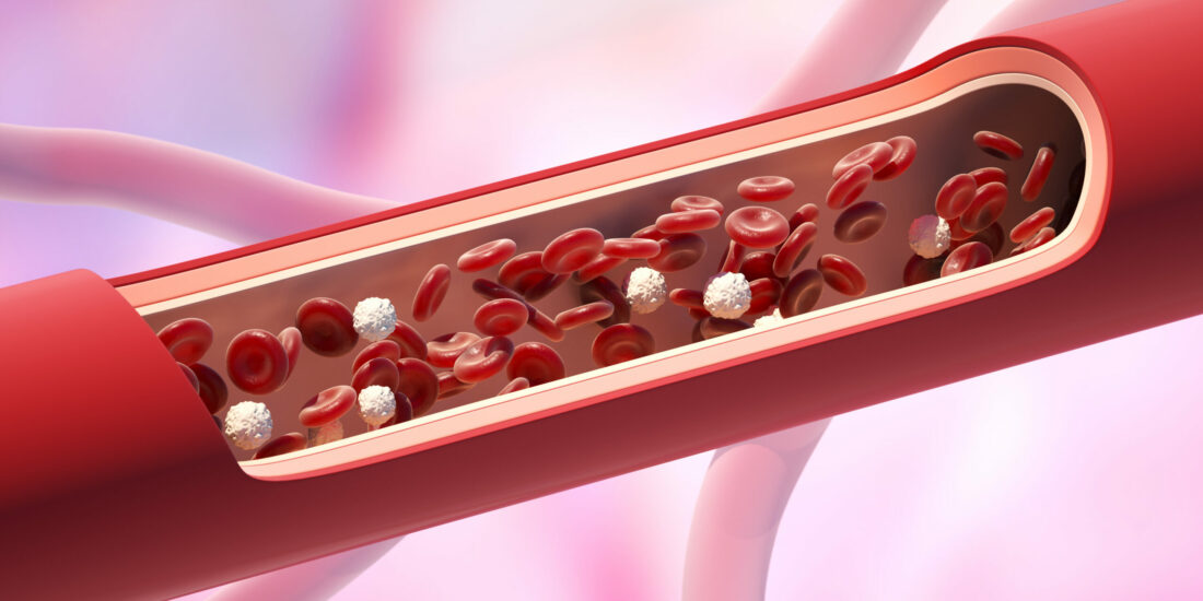
- Platelets are small, non-nucleated cells with a short lifespan (7–9 days) and specific structure (dense tubular system, different granules, lysosomes, mitochondria, and numerous adhesion molecules).
- Platelets regulate cell-cell interactions and local circulation through eicosanoids from arachidonic acid.
- Sigma-1 receptor ligands act at multiple points in arachidonic acid metabolism and play an important role in the control of the microcirculation by modulating the eicosanoid synthesis of the platelets and vessels.
- The effect of sigma-1 receptor ligands on platelet arachidonic acid metabolism is likely to occur not only through the modulation of COX and LOX but also through the modulation of specific enzymes.
- Sigma-1 receptor ligands may play an important role in the regulation of microcirculation by modulating platelet and vascular eicosanoid synthesis.
Abstract
Platelets regulate cell-cell interactions and local circulation through eicosanoids from arachidonic acid. Sigma non-opioid intracellular receptor 1 (sigma-1 receptor) expressed in platelets and endothelial cells can regulate intracellular signalization. Our aim was to examine the influence of sub-chronic, in vivo-administered sigma-1 receptor ligands 2-morpholin-4-ylethyl 1-phenylcyclohexane-1-carboxylate (PRE-084); N-benzyl-2-[(1S)-6,7-dimethoxy-1,2,3,4-tetrahydroisoquinolin-1-yl]ethan-1-amine; dihydrochloride, a new compound ((S)-L1); and N-[2-[4-methoxy-3-(2-phenylethoxy)phenyl]ethyl]-N-propylpropan-1-amine (NE-100) on the ex vivoarachidonic acid metabolism of the platelets and aorta of male rats. The serum level of sigma-1 receptor ligands was determined by liquid chromatography-mass spectrometry. Sigma-1 receptor and cyclooxygenase gene expression in the platelets were determined by a reverse transcription-coupled quantitative polymerase chain reaction. The eicosanoid synthesis was examined using a radiolabeled arachidonic acid substrate and enzyme-linked immunosorbent assay. We confirmed the absorption of sigma-1 receptor ligands and confirmed that the ligands were not present during the ex vivo studies, so their acute effect could be excluded. We detected no changes in either sigma-1 receptor or cyclooxygenase mRNA levels in the platelets. Nevertheless, (S)-L1 and NE-100 increased the quantity of cyclooxygenases there. Both platelet and aortic eicosanoid synthesis was modified by the ligands, although in different ways. The effect of the new sigma-1 receptor ligand, (S)-L1, was similar to that of PRE-084 in most of the parameters studied but was found to be more potent. Our results suggest that sigma-1 receptor ligands may act at multiple points in arachidonic acid metabolism and play an important role in the control of the microcirculation by modulating the eicosanoid synthesis of the platelets and vessels.
Graphical abstract

1. Introduction
Platelets are small, non-nucleated cells with a short lifespan (7–9 days) and specific structure (dense tubular system, different granules, lysosomes, mitochondria, and numerous adhesion molecules). This complex structure allows their involvement in hemostasis, cell-cell interaction (monocytes, endothelial cells, and lymphocytes), inflammatory processes, natural and acquired immunity, and regulation of the microcirculation (Gremmel et al., 2016; Koupenova et al., 2018; Mancuso and Santagostino, 2017a; van der Meijden and Heemskerk, 2019). Bioactive lipid mediators, i.e. eicosanoids (prostaglandins, thromboxane, and lipoxygenase products), synthesized by platelets participate in these processes (Yeung et al., 2018). Eicosanoids, which regulate cellular and vascular function, are generated from free arachidonic acid released from phospholipids in the cell membrane by phospholipase A2 in the presence of ionized calcium by cyclooxygenases (prostaglandin G/H synthases, EC1.14.99.1; COXs), lipoxygenases (EC1.13.11; LOXs), and specific synthases (Paes et al., 2019). Sigma non-opioid intracellular receptor 1 (the sigma-1 receptor), which modulates several cellular functions (Su et al., 2016; Aishwarya et al., 2021; Wang et al., 2021), is expressed on endothelial cells (Amer et al., 2013) and platelets (Váczi et al., 2021). The beneficial effects of sigma-1 receptor agonists have been described in different pathological conditions, such as sepsis (Rosen et al., 2019), ischemic-reperfusion injury (Gao et al., 2018), stroke (Nardai et al., 2020), and cardiovascular diseases (Tagashira et al., 2013). The expression of the sigma-1 receptor gene (Sigmar1) has been shown to reduce inflammatory cytokine production (Rosen et al., 2019) and to play a protective role against diabetic complications (Ha et al., 2012).
We aimed to investigate the effects of the sigma-1 receptor ligands on platelet and aortic eicosanoid synthesis in the rats. We hypothesized that sigma-1 receptor ligands administered sub-chronically in vivo (daily for one week) can modulate the ex vivo arachidonic acid metabolism of the platelets and aorta when the ligand is not currently present. For our studies, we chose two well-characterized ligands and one new sigma-1 receptor ligand. PRE-084 was initially identified as a selective agonist of the sigma-1 receptor (Su et al., 1991), which regulates cytokine production in stroke (Allahtavakoli and Jarrott, 2011), reduces microglial activation in traumatic brain injury, and has protective effects in neurogenic inflammation (Dong et al., 2016) and in the maintenance of the endothelial barrier (Motawe et al., 2020). NE-100 is an antagonist of the sigma-1 receptor (Okuyama and Nakazato, 1996), which has been shown to block sigma-1 receptor agonist (SA4503)–induced thoracic aortic vasodilation (Tagashira et al., 2013). The third ligand, (S)-L1, is a recently synthesized new compound, which binds with high affinity to the sigma-1 receptor (Dvorácskó et al., 2021). The serum level of the sigma-1 receptor ligands was determined by liquid chromatography-mass spectrometry (LC-MS) before the ex vivo analysis. The eicosanoid synthesis was examined using radiolabeled substrate and enzyme-linked immunosorbent assay. The effect of sigma-1 receptor ligands not only depends on the quantity and activity of the enzymes but can also be influenced by the levels of mRNA of genes coding the sigma-1 receptor and COX (Ptgs), which originate from megakaryocytes and are stored in the platelets. To confirm this process, the gene expression of the sigma-1 receptor and COXs in the platelets was also determined.
…
3. Results
3.1. Molecular modeling of the docking of the selected ligands to sigma non-opioid intracellular receptor 1
In a recent publication, we described a verified virtual screening protocol to identify novel sigma-1 receptor ligands. The sigma-1 and sigma-2 receptor–binding affinities of the highest-ranked compounds were measured in competitive radioligand binding. As a result of this study, we identified several new sigma-1 receptor ligands, among them compound (S)-L1 (N-benzyl-2-[(1S)-6,7-dimethoxy-1,2,3,4-tetrahydroisoquinolin-1-yl]ethan-1-amine; dihydrochloride, see Fig. 1A), which possesses high binding affinity (Ki = 11 ± 3 nM) to sigma-1 receptor and moderate sigma-1/sigma-2 receptor selectivity (Dvorácskó et al., 2021).

(A) Chemical structure of the sigma non-opioid intracellular receptor 1 ligands PRE-084, NE-100, and (S)-L1. (B) Binding pose of the agonist PRE-084 (gray), antagonist NE-100 (green), and (S)-L1 (yellow). The binding pose of NE-100 was extracted from the X-ray structure of the ligand-bound sigma non-opioid intracellular receptor 1 (PDB ID: 6DK0). The binding pose of the other two ligands was obtained with Glide XP docking to the apo receptor extracted from chain C of PDB structure 6DK1. To superimpose the binding pockets of 6DK0 and 6DK1, we used the backbone atoms of residues 170–176 and 197–217. (link to the animation sequence should be inserted here).
The chemical structure of PRE-084, NE-100, and (S)-L1 is presented in Fig. 1A. Every ligand fits the early pharmacophore model developed by Glennon (Glennon et al., 1994). It includes the following structural elements: a basic amine site that is necessary for the formation of an electrostatic bond with Glu172 of the receptor. It is typically flanked by two different hydrophobic groups. The hydrophobic parts are mostly aromatic and/or rigid heterocyclic sites. The binding modes of these ligands are compared in Fig. 1B. The binding pose of NE-100 was extracted from the X-ray structure of the ligand-bound sigma-1 receptor (PDB ID: 6DK0). The binding pose of the other two ligands was obtained with Glide XP docking (Schrödinger, 2019) to the apo receptor based on chain C of PDB structure 6DK1.
According to Schmidt et al. (2018), the main difference between agonist and antagonist binding is the presence of the interaction of the ligand with the C-terminal helix shown in the foreground of Fig. 1B. The cyclohexane ring of PRE-084 is the only moiety that can interact with this helix (Schmidt et al., 2018). The (S)-L1 and the sigma-1 receptor antagonist NE-100 have very similar binding poses (link to the animation sequence should be inserted here).
3.2. Physical and laboratory parameters of the animals
The significant increase in terminal body weight of the animals compared to basal levels correlates with the physiological, time-proportional increase in body weight. No significant difference in terminal body weights was observed between the sigma-1 receptor ligand-treated and the vehicle-treated rat groups. Age- and gender-matched, slightly elevated blood glucose levels in the animal groups are within the reference values published by Charles River Laboratories (Charles River, 2008) (Table 1).
At the end of the in vivo experiment (terminal phase), we detected no statistically significant difference in the platelet counts between the groups of animals (Table 1).
Table 1. Body weight, fasting serum glucose levels, and platelet number of the animals.
| Rats | Body weight (g) | Serum glucose (mM) | Platelet (109/L) | ||
|---|---|---|---|---|---|
| Basal | Terminal | Basal | Terminal | Terminal | |
| Vehicle | 364 ± 22 | 471 ± 31*** | 6.5 ± 0.2 | 7.3 ± 0.9 | 587 ± 86 |
| PRE-084 | 337 ± 27 | 445 ± 11*** | 6.4 ± 0.2 | 7.9 ± 0.4 | 685 ± 38 |
| (S)-L1 | 375 ± 21 | 460 ± 14*** | 6.7 ± 0.2 | 8.2 ± 0.4 | 589 ± 71 |
| NE-100 | 337 ± 20 | 466 ± 19*** | 6.8 ± 0.3 | 6.3 ± 0.3 | 713 ± 137 |
Data is shown as mean ± SD; n = 9–9 rats/group. We determined the basal body weights and fasting (12 h) blood glucose levels of the animals injected with the vehicle or sigma-1 receptor ligands. ANOVA, Bonferroni test, ***p < 0.001 terminal body weights were compared to their own basal body weights.
3.3. Concentration of the sigma non-opioid intracellular receptor 1 ligands in rat plasma
3.3.1. In the preliminary experiments
Prior to the in vivo administration of the sigma-1 receptor ligands, we monitored the kinetic changes in the plasma levels of PRE-084, (S)-L1, and NE-100 administered intraperitoneally as a single dose in the male rats (Supplementary Figs. S1–S3). A significant elevation in plasma levels was detected one hour after the administration of PRE-084 and 30 minutes after the administration of (S)-L1 and NE-100. The ligand concentration rapidly decreased, and the quantity of sigma-1 receptor ligands was insignificant or undetectable 20 hours after the injection.
3.3.2. In the terminal phase of the in vivo experiment
Plasma concentrations of the sigma-1 receptor ligands were determined in all of the rats 20 hours after the last ligand injection immediately before the ex vivo studies. The plasma concentrations of the sigma-1 receptor ligands were below the limit of detection or quantification 20 hours after the last dose (Table 2).
Table 2. Concentration of the sigma non-opioid intracellular receptor 1 ligands in rat plasma. Samples were taken 20 hours after the last dose (3 mg/bwkg/day for seven days).
| Analyte | Concentration (M) |
|---|---|
| PRE-084 | < LOQ |
| (S)-L1 | 5.64 ± 0.32 |
| NE-100 | < LOD |
Data is shown as mean ± SD; n = 9–9 rats/group; LOQ: limit of quantification; LOD: limit of detection.
3.4. Effects of the sigma non-opioid intracellular receptor 1 ligands on the Sigmar1 mRNA level in the rat platelets
The mRNA levels of Sigmar1 were detected in the rat platelet samples using RT-qPCR. The Sigmar1 mRNA showed a low concentration level in the rat platelets. Although the Ct values were high in most of the cases, we considered them reliable because the RT-qPCR was performed with TaqMan assays. The daily administration of the sigma-1 receptor ligands for one week did not change the level of Sigmar1mRNA in the platelets (Fig. 2).

Relative normalized transcript levels of Sigmar1 in the platelet samples from the rats treated with the sigma non-opioid intracellular receptor 1 ligands. The rats were treated with 3 mg/bwkg/day PRE-084, (S)-L1, or NE-100 for seven days. The vehicle-treated group received saline solution. The transcript levels of Sigmar1 in the rat platelet samples were first normalized to their own endogenous control mRNA levels (Gapdh) and then to the similarly normalized mRNA levels in the vehicle-treated animal groups. Fold changes were calculated using the 2−ΔΔCt formula. Mean ± SD, n = 3 samples pooled from nine rats, ANOVA, Dunnett test.
3.5. Analysis of the ex vivo arachidonic acid metabolism in the rats
3.5.1. Effects of the sigma non-opioid intracellular receptor 1 ligands on the ex vivo eicosanoid synthesis of the platelets
3.5.1.1. Application of radioactive arachidonic acid substrate to study platelet eicosanoid synthesis
The total quantity of the radioactive cyclooxygenase metabolites (COX; sum of 6-keto prostaglandin F1α/6-k-PGF1α, which is a stable metabolite of prostacyclin; prostaglandin F2α/PGF2α; prostaglandin E2/PGE2; prostaglandin D2/PGD2; thromboxane B2/TxB2, which is a stable metabolite of TxA2; and 12-L-hydroxy-5,8,10-heptadecatrienoic acid/12-HHT) in the rat platelets was significantly reduced by both PRE-084 and (S)-L1, while it was elevated by NE-100 (Table 3).
While the total quantity of radioactive LOX metabolites was raised by PRE-084 and (S)-L1, it was lowered by NE-100, thus indicating an opposite effect on the total quantity of radioactive COX and LOX metabolites. The sigma-1 receptor ligands did not change the total quantity of radioactive arachidonic acid metabolites (COX + LOX). The COX/LOX ratio was significantly reduced in the PRE-084 or (S)-L1 groups, while it was increased by NE-100 compared to the vehicle-treated rat platelets (Table 3).
Table 3. Arachidonic acid metabolism in the rat platelets.
| RATS | COX (cpm) | LOX (cpm) | COX + LOX (cpm) | COX/LOX |
|---|---|---|---|---|
| Vehicle | 1425 ± 63 | 3132 ± 291 | 4649 ± 137 | 0.44 ± 0.01 |
| PRE-084 | 1271 ± 84* | 3437 ± 100** | 4726 ± 51 | 0.37 ± 0.01*** |
| (S)-L1 | 1146 ± 84*** | 3428 ± 122** | 4568 ± 170 | 0.33 ± 0.01*** |
| NE-100 | 1897 ± 76*** | 2788 ± 111*** | 4685 ± 93 | 0.68 ± 0.02*** |
cpm: count per minute; COX: total quantity of radioactive cyclooxygenase metabolites synthesized from arachidonic acid in platelets; LOX: total quantity of radioactive lipoxygenase metabolites synthesized from arachidonic acid in platelets. All data are represented as mean ± SD. n = 9–9 rats/group. ANOVA, Bonferroni test, *p < 0.05, **p < 0.03, ***p < 0.01 compared to the vehicle-treated groups.
In the platelets, the synthesis of TxB2 (stable metabolite of TxA2, the main vasoconstrictor and platelet aggregator product) and the production of PGD2(vasodilator and platelet anti-aggregator metabolite) were significantly decreased in the PRE-084 and (S)-L1 groups, while they were increased in the NE-100 group as compared to the vehicle-treated samples (Fig. 3).

The effects of the sigma non-opioid intracellular receptor 1 ligands on the different radioactive cyclooxygenase metabolites in the rat platelets. The y-axis represents the isotope activity in count per minute (cpm). All data are represented as mean ± SD. n = 9–9 rats/group. ANOVA, Bonferroni test, **p < 0.03, ***p < 0.01 compared to the vehicle-treated groups.
In the platelets, the quantity of vasoconstrictor and platelet aggregator radioactive cyclooxygenase metabolites (CON: the sum of PGF2α and TxB2) was reduced by PRE-084 and (S)-L1 but raised by NE-100 (Table 4). The quantity of vasodilator and anti-aggregator radioactive cyclooxygenase metabolites (DIL: the sum of PGE2 and PGD2) was lowered by PRE-084, but it was boosted by NE-100. We can see a significant reduction in the ratio of the CON and DIL metabolites in the (S)-L1 ligand group (Table 4).
Table 4. Vasoconstrictor, platelet aggregator (CON) and vasodilator, platelet anti-aggregator (DIL) radioactive COX metabolites in the rat platelets.
| RATS | CON (cpm) | DIL (cpm) | CON/DIL |
|---|---|---|---|
| Vehicle | 840 ± 34 | 354 ± 26 | 2.43 ± 0.21 |
| PRE-084 | 763 ± 66** | 319 ± 26** | 2.37 ± 0.12 |
| (S)-L1 | 478 ± 71*** | 356 ± 80 | 1.41 ± 0.43*** |
| NE-100 | 1103 ± 100*** | 356 ± 80 | 2.19 ± 0.3 |
cpm: count per minute; CON: the sum of PGF2α and TxB2; DIL: the sum of PGE2 and PGD2. All data are represented as mean ± SD. n = 9–9 rats/group. ANOVA, Bonferroni test, **p < 0.03, ***p < 0.01 compared to the vehicle-treated groups.
3.5.1.2. Cyclooxygenase enzyme levels in the platelets detected by ELISA
We detected both COX-1 and COX-2 enzymes in the platelets by ELISA. The concentration of COX-2 in the rat platelets was higher than that of COX-1. The level of the COX-1 and COX-2 enzymes was elevated by all of the sigma-1 receptor ligands used but in a different manner. The largest total concentration of these enzymes (COX-1+COX-2) was detected in the (S)-L1 ligand group (Table 5).
Table 5. Effect of the sigma non-opioid intracellular receptor 1 ligands on the quantity of COX-1 and COX-2 enzymes in the rat platelets determined by ELISA.
| RATS | COX-1 | COX-2 | COX-1+COX-2 | COX-1/COX-2 |
|---|---|---|---|---|
| (ng/mL) | (ng/mL) | (ng/mL) | Empty Cell | |
| Vehicle | 4.37 ± 1.3 | 5.89 ± 1.9 | 10.59 ± 3.3 | 0.77 ± 0.14 |
| PRE-084 | 5.5 ± 1.1 | 6.99 ± 2.2 | 12.87 ± 3.0 | 0.72 ± 0.09 |
| (S)-L1 | 6.17 ± 0.9** | 11.52 ± 0.9*** | 17.89 ± 1.0*** | 0.51 ± 0.09** |
| NE-100 | 6.39 ± 0.8*** | 10.43 ± 1.33*** | 16.81 ± 2.0*** | 0.62 ± 0.06* |
COX-1: type 1 cyclooxygenase; COX-2: type 2 cyclooxygenase. All data are represented as mean ± SD. n = 9–9 rats/group. ANOVA, Bonferroni test, *p < 0.05, **p < 0.03, ***p < 0.01 compared to the vehicle-treated groups
3.5.2. Effects of sigma non-opioid intracellular receptor 1 ligands on the ex vivo eicosanoid synthesis of the aorta
3.5.2.1. Application of the radioactive arachidonic acid substrate for the study of aortic eicosanoid synthesis
The quantity of radioactive COX metabolites in the rat aorta was significantly reduced by PRE-084, while the quantity of radioactive LOX products was significantly lower in the (S)-L1 group, compared to the vehicle-treated group (Table 6).
Table 6. Arachidonic acid metabolism in the rat abdominal aorta.
| RATS | COX (cpm) | LOX (cpm) | COX + LOX (cpm) | COX/LOX |
|---|---|---|---|---|
| Vehicle | 915 ± 215 | 551 ± 178 | 1466 ± 346 | 1.75 ± 0.4 |
| PRE-084 | 717 ± 157* | 421 ± 87 | 1094 ± 153** | 1.8 ± 0.7 |
| (S)-L1 | 826 ± 133 | 305 ± 67*** | 1132 ± 137* | 2.86 ± 0.8*** |
| NE-100 | 1443 ± 202*** | 833 ± 51*** | 2277 ± 247*** | 1.73 ± 0.2 |
cpm: count per minute; COX: total quantity of radioactive cyclooxygenase metabolites synthesized from arachidonic acid in the abdominal aorta; LOX: total quantity of radioactive lipoxygenase metabolites synthesized from arachidonic acid in the abdominal aorta. All data are represented as mean ± SD. n = 9–9 rats/group. ANOVA, Bonferroni test, *p < 0.05, **p < 0.03, ***p < 0.01 compared to the vehicle-treated groups.
The quantity of radioactive COX and LOX metabolites in the rat aorta was significantly elevated in the NE-100 group compared to the vehicle-treated samples. The total quantity of radioactive arachidonic acid metabolites (COX + LOX) was significantly lower in the PRE-084 and (S)-L1 groups but higher in the NE-100 sigma-1 receptor antagonist group compared to the vehicle-treated rat aorta. The COX/LOX ratio of the radioactive arachidonic acid metabolites in the rat aorta significantly increased in the (S)-L1 group compared to the vehicle-treated animals (Table 6).
In the rat aorta, the synthesis of 6-k-PGF1α (stable metabolite of prostacyclin and the main vasodilator and platelet anti-aggregator product of the aorta) was significantly increased by (S)-L1 and NE-100 and reduced by PRE-084, compared to the vehicle-treated sample. The production of TxB2 in the rat aorta was significantly lowered by both PRE-084 and (S)-L1 but raised by NE-100 (Fig. 4).

The effects of the sigma non-opioid intracellular receptor 1 ligands on the different radioactive cyclooxygenase metabolites in the rat aorta. The y-axis represents the isotope activity in count per minute (cpm); 6-k-PGF1α: 6-keto prostaglandin F1α; TxB2: thromboxane B2. All data are represented as mean ± SD. n = 9–9 rats/group. ANOVA, Bonferroni test, *p < 0.05, **p < 0.03, ***p < 0.01 compared to the vehicle-treated groups.
PRE-084 significantly decreased the quantity of DIL radioactive COX metabolites, while NE-100 increased the quantity of both DIL and CON radioactive COX metabolites in the rat aorta compared to the vehicle-treated group. Overall, NE-100 lowered the CON/DIL ratio of radioactive COX metabolites compared to the vehicle-treated group (Table 7).
Table 7. Vasoconstrictor, platelet aggregator (CON) and vasodilator, platelet anti-aggregator (DIL) radioactive COX metabolites in the rat aorta.
| RATS | CON (cpm) | DIL (cpm) | CON/DIL |
|---|---|---|---|
| Vehicle | 539 ± 128 | 242 ± 28 | 2.22 ± 0.4 |
| PRE-084 | 459 ± 115 | 206 ± 14*** | 2.28 ± 0.59 |
| (S)-L1 | 533 ± 93 | 236 ± 35 | 2.30 ± 0.44 |
| NE-100 | 813 ± 139*** | 477 ± 74*** | 1.72 ± 0.24*** |
cpm: count per minute; CON: the sum of PGF2α and TxB2; DIL: the sum of 6-k-PGF1α (which is a stable metabolite of prostacyclin), PGE2, and PGD2. All data are represented as mean ± SD. n = 9–9 rats/group. ANOVA, Bonferroni test, *p < 0.05, ***p < 0.01 compared to the vehicle-treated groups.
3.5.2.2. Cyclooxygenase enzyme levels in the aorta measured by ELISA
We detected a higher concentration of COX-2 than COX-1 (Table 8) in the rat aorta.
Table 8. Effect of sigma non-opioid intracellular receptor 1 ligands on the COX-1 and COX-2 enzyme levels in the rat aorta as determined by ELISA.
| RATS | COX-1 | COX-2 | COX-1+COX-2 | COX-1/COX-2 |
|---|---|---|---|---|
| (ng/mL) | (ng/mL) | (ng/mL) | Empty Cell | |
| Vehicle | 3.70 ± 1.0 | 9.1 ± 2.1 | 13.27 ± 1.88 | 0.39 ± 0.14 |
| PRE-084 | 5.84 ± 1.1*** | 10.84 ± 0.41 | 16.66 ± 1.65 | 0.54 ± 0.08 |
| (S)-L1 | 8.41 ± 0.7*** | 13.86 ± 0.9*** | 21.89 ± 0.97*** | 0.61 ± 0.09* |
| NE-100 | 4.37 ± 0.9 | 5.49 ± 0.75*** | 10.31 ± 0.14 | 0.76 ± 0.03 |
COX-1: type 1 cyclooxygenase; COX-2: type 2 cyclooxygenase. All data are represented as mean ± SD. n = 9–9 rats/group. ANOVA, Bonferroni test, *p < 0.05, ***p < 0.01 compared to the vehicle-treated groups.
PRE-084 and (S)-L1 increased the aortic COX-1 and COX-2 concentrations, with a higher elevation observed for COX-1 (Table 8). NE-100 reduced the COX-2 level, therefore, and shifted the balance between the enzymes to COX-1. The most effective sigma-1 receptor ligand was (S)-L1, which increased the level of COX-1 and COX-2 (Table 8).
3.6. Effects of sigma non-opioid intracellular receptor 1 ligands on the Ptgs1 mRNA level in the rat platelets
The daily administration of sigma-1 receptor ligands for one week did not change the level of Ptgs1 mRNA in the rat platelets (Fig. 5). Interestingly, the effect of (S)-L1 was different in the case of the Sigmar1 and Ptgs1 mRNA levels: an effect similar to the sigma-1 receptor agonist PRE-084 was observed for the Sigmar1 mRNA level, while a similar effect of the sigma-1 receptor antagonist NE-100 was seen for the Ptgs1 mRNA concentration in the rat platelets. We measured but could not detect Ptgs2 mRNA by RT-qPCR in the platelet samples using 40 cycles.

Relative normalized transcript levels of Ptgs1 in the rat platelet samples treated with sigma non-opioid intracellular receptor 1 ligands. The rats were treated with 3 mg/bwkg/day PRE-084, (S)-L1, or NE-100 for seven days. The vehicle-treated group received only saline solution. The transcript levels of Ptgs1 in the rat platelet samples were first normalized to their own endogenous control mRNA levels (Gapdh) and then to the similarly normalized mRNA levels in the vehicle-treated animal groups. Fold changes were calculated using the 2−ΔΔCtformula. Mean ± SD, n = 3 samples pooled from nine rats, ANOVA, Dunnett test.
4. Discussion
In this study, we investigated the effects of sub-chronic, in vivo-administered sigma-1 receptor ligands on platelet and aortic eicosanoid synthesis in healthy male rats. For our studies, we selected the sigma-1 receptor ligands PRE-084, NE-100, and (S)-L1 based on their binding strength to the receptor and their binding position in the sigma-1 receptor binding pocket (Fig. 1). Treatment duration was determined by platelet lifespan (one week) (Thon and Italiano, 2012; Cimmino and Golino, 2013). We confirmed that all the sigma-1 receptor ligands administered intraperitoneally entered the circulation, but their 30 min serum levels showed a large variation, which may indicate a difference in their absorption rate. The difference observed in the time-dependent analysis of the serum levels of the ligands (Figs. S1–S3) could be explained by their distinct metabolism and excretion. None of the ligands we studied at the dose used in our studies and for the duration involved caused metabolic changes in the rats that resulted in a change in body weight or blood glucose levels (over the time period). No effect of sub-chronic treatment was observed on platelet formation with sigma-1 receptor ligands, although this is not relevant in the present study, as their eicosanoid synthesis was monitored at standard platelet counts (2.5 × 10⁸ platelets/μL) (Table 1).
In order to rule out the direct effects of sigma-1 receptor ligands, our ex vivo/in vitrostudies were started 20 h after the last ligand treatment, when the serum levels of ligands were already below the limit of detection or quantification (Table 2). Thus, the changes in arachidonic acid metabolism induced by the sigma-1 receptor ligands in the platelets and aorta and detected ex vivo may be due to changes in platelet and aortic function induced by the ligands in vivo. The sigma-1 receptor ligands we tested also affected rat platelet and aortic arachidonic acid metabolism in different ways (Table 3, Table 6).
In the rat platelets, none of the ligands induced changes in either sigma-1 receptor or COX mRNA levels; that is, the difference in arachidonic acid metabolism cannot be explained by the effects of the ligands on either sigma-1 receptor or cyclooxygenase enzyme transcription (Fig. 2, Fig. 5). No change in COX enzyme concentration was detected in the rat platelets by ELISA after in vivo treatment with the PRE-084 ligand (Table 5). Therefore, a decrease in the total quantity of arachidonic acid metabolites formed by the cyclooxygenase pathway may be explained by inhibition of the COX enzyme or by a decrease in the absolute or relative quantity of free arachidonic acid substrate (Table 3). The generation of arachidonic acid metabolites is dependent on the presence of sufficient quantity of free arachidonic acid, which may be influenced by membrane phospholipid composition, phospholipase activity, and [Ca2+]i levels. Sigma-1 receptor is known to be able to alter all these factors (Hayashi, 2019; Paes et al., 2019; Penke et al., 2018; Starr and Werling, 2002). No drop in the total quantity of arachidonic acid metabolites (COX + LOX) was observed with PRE-084, so an absolute reduction in free arachidonic acid due to higher phospholipid re-acylation can be ruled out. The decrease in the total quantity of COX metabolites could have been caused by a relative substrate decrease due to direct inhibition of cyclooxygenase and/or increased lipoxygenase activity, as supported by the drop in the COX/LOX ratio (Table 3). While no data are available for the direct action of sigma-1 receptor ligands on COX activity, a recent lipidomics study found a downregulation of the COX pathway due to bufotenin, a sigma-1 receptor ligand (Wang et al., 2021).
In contrast to the PRE-084 ligand, both (S)-L1 and NE-100 induced a similar increase in COX enzyme levels in the rat platelets (Table 5). However, (S)-L1 reduced the formation of COX metabolites, whereas NE-100 boosted it (Table 3). The higher COX enzyme levels and lower COX metabolite levels suggest that the PRE-084 and (S)-L1 ligands may have different effects on the translation and synthesis of COX enzymes, as well as on enzyme activity and metabolite production. The effects of (S)-L1 and NE-100 were the opposite not only on COX product formation but also on that of LOX (Table 3). In parallel with the elevation of the arachidonic acid substrate, the expression and/or activity of COX enzymes might be stimulated. Of the two COX isoforms, COX-1 is constitutively expressed in various tissues, but the expression of COX-2 is mostly inducible. Although the platelets are non-nucleated cells, they can de novo synthesize COX-1 from the cytoplasmic mRNA that originates from the megakaryocyte (Evangelista et al., 2006). In a previous report, COX-2 mRNA and protein were detected in human platelets, but in a significantly smaller quantity than COX-1 (Hu et al., 2017). However, under our present experimental conditions, we were unable to detect COX-2 (Ptgs2) mRNA in rat platelets by 40-cycle RT-qPCR. This may be explained by the limited pool of mRNA transcripts from megakaryocytes that can be used for protein (e.g. COX-2) synthesis, so increased arachidonic acid metabolism may lead to depletion of intracellular reserves (mRNA, enzyme pool, and [Ca2+]i).
The effect of sigma-1 receptor ligands on platelet arachidonic acid metabolism is likely to occur not only through the modulation of COX and LOX but also through the modulation of specific enzymes. This is suggested by the fact that the administration of (S)-L1 in the rats shifted the ratio of CON/DIL COX products in the platelets (Table 4). The drop in the CON/DIL ratio in the platelets was induced by (S)-L1 by lowering the formation of CON products (Table 4 and Fig. 3). The synthesis of the main platelet cyclooxygenase metabolite, the vasoconstrictor- and platelet-aggregating thromboxane, was reduced much more by our new ligand, (S)-L1, than by PRE-084 (Fig. 3).
Under physiological conditions, platelet and endothelial cell arachidonic acid metabolism is known to differ significantly. Platelets primarily produce vasoconstrictor thromboxane, which induces platelet aggregation, and the endothelium primarily produces vasodilator prostacyclin, which inhibits it; however, under physiological conditions, these products are in balance and thus ensure physiological microcirculation (Csányi et al., 2007; Mezei et al., 2017; Mitchell and Kirkby, 2019).
The effects of the sigma-1 receptor ligands we studied on arachidonic acid metabolism in the rat aorta showed a significant difference compared to their effects on platelets. In contrast to platelets, the total quantity of eicosanoids formed by the aorta via the lipoxygenase pathway was decreased by (S)-L1, whereas NE-100 increased it. Sigma-1 receptor ligands induced a change in the aorta that was in the same in direction as that in the platelets but smaller in total quantity of COX metabolites (Table 3, Table 6). These changes can be explained by changes in both absolute and relative arachidonic acid substrate quantity. The drop in the total quantity of arachidonic acid metabolites (COX + LOX) by (S)-L1 and the increase due to NE-100 suggest that sigma-1 receptor ligands may have affected arachidonic acid release from phospholipids, i.e. the absolute quantity of substrate. On the other hand, the shift in the ratio of cyclooxygenase to lipoxygenase metabolites (COX/LOX) in the aorta due to (S)-L1 may suggest that the ligand affected the two enzyme systems differentially, resulting in a relative substrate deficiency (Table 6). Although the effects of the ligands on the total quantity of platelet and aortic cyclooxygenase metabolites were similar (Table 3, Table 6), their effects on the ratio of cyclooxygenase-derived vasoconstrictor and platelet aggregator (CON) and vasodilator, platelet aggregation inhibitor cyclooxygenase metabolites (DIL) (CON/DIL) were significantly different. (S)-L1 in the platelets and NE-100 in the aorta resulted in a decrease in CON/DIL, whereas the other ligands produced no change (Table 4, Table 7; Fig. 3, Fig. 4). NE-100 lowered the CON/DIL ratio in the aorta despite raising the synthesis of both CON and DIL metabolites (Table 7 and Fig. 4). The synthesis of the major aortic cyclooxygenase metabolite, the vasodilator and anti-aggregator prostacyclin, was enhanced by the new ligand we tested, (S)-L1, which was inhibited by PRE-084 (Fig. 4).
The neuroprotective (Penke et al., 2018) and cardioprotective (Lewis et al., 2020) effects of the sigma-1 receptor have already been demonstrated. However, only a fraction of all cardiovascular diseases is of cardiac origin, and macro- and microvascular dysfunction and/or platelet activation are responsible for the development and exacerbation of many diseases. The eicosanoids are de novo, locally formed (Evangelista et al., 2006), short half-life (Egan and FitzGerald, 2006; Paes et al., 2019), autocrine-, paracrine-, and endocrine-acting (Paes et al., 2019) arachidonic acid metabolites. Under physiological conditions, the balance of vasoactive and platelet aggregation-mediated cyclooxygenase products synthesized by platelets and vascular endothelium ensures proper microcirculation and tissue perfusion (Mancuso and Santagostino, 2017a; Mitchell and Kirkby, 2019). The sub-chronic in vivo administration of the PRE-084, (S)-L1, and NE-100 sigma-1 receptor ligands in healthy rats modulated both rat platelet and aortic eicosanoid synthesis; however, these effects were cell/tissue-specific. On platelet and aortic eicosanoid synthesis, the effects of our ligands resulted in a predominance of vasodilator and platelet aggregation inhibitory COX eicosanoids. Under our present study conditions, despite the different mode and intensity of action, all three ligands we investigated played a protective role in maintaining the balance of platelet and vascular eicosanoid synthesis, i.e. tissue perfusion and microcirculation. Of the sigma-1 receptor ligands we tested, (S)-L1 proved to be the most potent.
5. Conclusion
Our results suggest that sigma-1 receptor ligands may act at multiple points in arachidonic acid metabolism. This effect was found to be cell-specific. Our experimental results on eicosanoid synthesis in rat platelets confirm the regulatory role of the sigma-1 receptor in cellular functions.
(S)-L1, a readily absorbed and rapidly eliminated/metabolized novel sigma-1 receptor ligand, affected rat platelet and aortic arachidonic acid metabolism in a manner similar to the sigma-1 receptor agonist PRE-084, but in a manner different from the antagonist NE-100.
Sigma-1 receptor ligands may play an important role in the regulation of microcirculation by modulating platelet and vascular eicosanoid synthesis.