
Blockade of the Sigma-1 Receptor Relieves Cognitive and Emotional Impairments Associated to Chronic Osteoarthritis Pain

By Mireia Carcolé, Daniel Zamanillo, Manuel Merlos, Begoña Fernández-Pastor, David Cabañero, and Rafael Maldonado
Excerpt from the article published in Frontiers in Pharmacology, 03 May 2019
Sec. Experimental Pharmacology and Drug Discovery DOI: https://doi.org/10.3389/fphar.2019.00468
Abstract
Osteoarthritis is the most common musculoskeletal disease worldwide, often characterized by degradation of the articular cartilage, chronic joint pain and disability. Cognitive dysfunction, anxiety and depression are common comorbidities that impact the quality of life of these patients. In this study, we evaluated the involvement of sigma-1 receptor (σ1R) on the nociceptive, cognitive and emotional alterations associated with chronic osteoarthritis pain. Monosodium iodoacetate (MIA) was injected into the knee of Swiss-albino CD1 mice to induce osteoarthritis pain, which then received a repeated treatment with the σ1R antagonist E-52862 or its vehicle. Nociceptive responses and motor performance were assessed with the von Frey and the Catwalk gait tests. Cognitive alterations were evaluated using the novel object recognition task, anxiety-like behavior with the elevated plus maze and the zero-maze tests, whereas depressive-like responses were determined using the forced swimming test. We also studied the local effect of the σ1R antagonist on cartilage degradation, and its central effects on microglial reactivity in the medial prefrontal cortex. MIA induced mechanical allodynia and gait abnormalities that were prevented by the chronic treatment with the σ1R antagonist. E-52862 also reduced the memory impairment and the depressive-like behavior associated to osteoarthritis pain. Interestingly, the effect of E-52862 on depressive-like behavior was not accompanied by a modification of anxiety-like behavior. The pain-relieving effects of the σ1R antagonist were not due to a local effect on the articular cartilage, since E-52862 treatment did not modify the histological alterations of the knee joints. However, E-52862 induced central effects revealed by a reduction of the cortical microgliosis observed in mice with osteoarthritis pain. These findings show that σ1R antagonism inhibits mechanical hypersensitivity, cognitive deficits and depressive-like states associated with osteoarthritis pain in mice. These effects are associated with central modulation of glial activity but are unrelated to changes in cartilage degradation. Therefore, targeting the σ1R with E-52862 represents a promising pharmacological approach with effects on multiple aspects of chronic osteoarthritis pain that may go beyond the strict inhibition of nociception.
Introduction
Osteoarthritis is one of the most prevalent chronic diseases and represents a major socio-economic burden worldwide (Johnson and Hunter, 2014; Puig-Junoy and Ruiz Zamora, 2015). It is a complex disease of the whole joint defined by progressive destruction of articular cartilage (Sutton et al., 2009; Zhang et al., 2013). Its most problematic symptoms are pain and loss of joint function, and current pharmacological therapies are limited and generally directed to relief pain. However, osteoarthritis pain is frequently accompanied by co-morbid affective manifestations, such as anxiety and depression (Axford et al., 2010; Goldenberg, 2010; Sharma et al., 2016), and by cognitive alterations including memory dysfunction, which contribute to an overall impairment of the quality of life (Moriarty et al., 2011; Moriarty and Finn, 2014). These co-morbid alterations could in turn aggravate pain perception and contribute to the establishment of chronic osteoarthritis pain (Villemure and Bushnell, 2009). In this context, treatments that simultaneously control the nociceptive, affective and cognitive manifestations could represent an efficient therapeutical approach for chronic osteoarthritis pain.
Sigma-1 receptor (σ1R) is a ligand-regulated chaperone that interacts with a large number of receptors and ion channels (Su and Hayashi, 2003; Hayashi and Su, 2007; Tsai et al., 2009) and has widespread distribution in the nervous system (Harada et al., 1994; Alonso et al., 2000; Kitaichi et al., 2000). Preclinical studies have implicated this receptor in several neurological disorders, such as addiction (Matsumoto et al., 2002; Maurice et al., 2002), schizophrenia (Hayashi et al., 2011) neurodegenerative disorders (Maurice et al., 1998; Francardo et al., 2014) or depression (Urani et al., 2001; Skuza and Rogóz, 2003; Lucas et al., 2008). σ1R has also been proposed as an effective therapeutic target in several models of chronic pain (Entrena et al., 2009; Nieto et al., 2012; Romero et al., 2012; Gris et al., 2014; Tejada et al., 2014). However, these studies do not assess the participation of the σ1R on the emotional or cognitive alterations that can develop after the induction of persistent pain (La Porta et al., 2015, 2016; Negrete et al., 2017). Thus, it remains to be determined whether σ1R ligands could be effective relieving chronic osteoarthritis pain together with its co-morbid cognitive and affective impairments.
The prefrontal cortex plays a crucial role in emotional processing (Gusnard et al., 2001; Etkin et al., 2011), cognitive functions (Phelps et al., 2004) and modulation of pain perception (Apkarian et al., 2004, 2005; Metz et al., 2009). Clinical studies have observed functional and structural abnormalities in the prefrontal cortex of patients suffering from chronic pain (Apkarian et al., 2004; Seminowicz et al., 2011). Such anatomical alterations have also been observed in animal models of neuropathic pain, where a decreased volume of the prefrontal cortex was found in correlation with anxiety-like behavior (Seminowicz et al., 2009). Several studies have also revealed the important role of microglial cells in the adaptative changes occurring in the central nervous system during chronic pain, leading to the persistence of pain manifestations (Racz et al., 2008). The role of microglia on chronic pain has been revealed in the spinal cord (Racz et al., 2008), and supraspinal activation of microglia is also partly responsible for the structural, functional, and molecular neuroplasticity associated with pathological pain (Boadas-Vaello et al., 2017). In fact, it has been proposed that microglial alterations in cortical regions underlie pain-induced emotional and cognitive impairments (Panigada and Gosselin, 2011). Therefore, targeting microglial reactivity in these areas could be an appropriate strategy to treat the affective and memory disturbances observed in chronic pain conditions. Interestingly, σ1R is highly expressed in microglia (Gekker et al., 2006) where it exerts a modulatory function (Peviani et al., 2014; Moritz et al., 2015). Therefore, it would be important to elucidate the possible role of σ1R on cortical microgliosis associated to chronic osteoarthritis pain.
Here we assessed the effect of the σ1R antagonist E-52862, also named S1RA (Romero et al., 2012; Gris et al., 2014) and MR309 (Castany et al., 2018), on the nociceptive, cognitive and emotional alterations observed in the monosodium iodoacetate (MIA) model of osteoarthritis pain in mice. To determine whether E-52862 exerts its effects through a local participation of σ1R on the knee joint, we analyzed levels of cartilage degradation through histological assessment. In addition, we evaluated possible central neuroplastic effects of the σ1R antagonist by determining microglial density and morphology in the medial prefrontal cortex.
…
Results
The σ1R Antagonist E-52862 Reverses Mechanical Hypersensitivity Associated to Osteoarthritis Pain
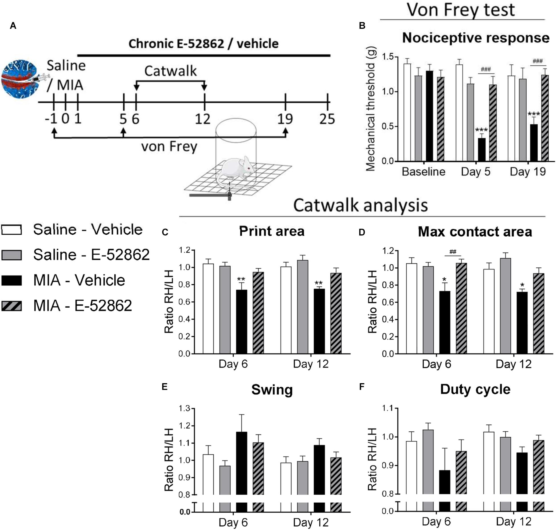
To evaluate the effect of E-52862 on mechanical hypersensitivity associated to osteoarthritis pain, mice were intraperitoneally treated twice a day with either vehicle or E-52862 (20 mg/kg) from the 1st day after MIA injection until the end of the experiment (day 25). Von Frey test was performed before and 5 and 19 days after MIA injection, and gait analysis was evaluated at 6 and 12 days (Figure 1A). MIA injection induced a persistent mechanical hypersensitivity in vehicle-treated mice (p < 0.001 vs. saline mice, days 5 and 19). Conversely, this decrease in mechanical thresholds was absent in mice treated with E-52862 (p < 0.001 vs. MIA vehicle mice, days 5 and 19) (Figure 1B). Gait analysis also showed MIA-induced alterations on walking patterns that were partly reversed by E-52862. Mice injected with MIA and treated with vehicle showed a significant decrease of the print area (p< 0.01 vs. saline; Figure 1C) and maximal contact area (p < 0.05 vs. saline; Figure 1D) at both time points tested. These alterations were not observed when MIA-injected mice were treated with E-52862 (Figures 1C,D). No significant effects were observed in the swing for any of the experimental groups (Figure 1E), however, a trend toward a decreased duty cycle was observed in MIA mice treated with vehicle (p = 0.08 vs. saline; Figure 1F). Therefore, blocking the σ1R produced a relief of mechanical pain associated to the injection of MIA that was also reflected into a normalization of gait function.
Repeated treatment with E-52862 reversed mechanical hypersensitivity associated to osteoarthritis pain.
(A) Mice received an intra-knee injection of MIA or saline and were treated with vehicle or E-52862 (20 mg/kg) from day 1 until the end of the experiment (day 25). Mechanical thresholds were assessed with the von Frey under basal conditions and on days 5 and 19 after the intra-articular injection, and gait was analyzed with the Catwalk test on days 6 and 12. (B) MIA-injected mice treated with vehicle showed a decrease on mechanical thresholds that was reversed in E-52862-treated mice. Catwalk analysis revealed a decrease of the ratio (right hind/left hind paws) of print area (C) and maximal contact area (D) in mice injected with MIA and treated with vehicle. This alteration was reversed in mice receiving E-52862. Swing (E) and duty cycle (F) were not significantly altered by the intra-knee injection. Data are expressed as mean ± SEM (n = 6–8 animals per group). ∗p < 0.05, ∗∗p< 0.01, ∗∗∗p < 0.001 vs. Saline-vehicle, ##p < 0.01, ###p < 0.001 for MIA-vehicle vs. MIA-E-52862 (3-way repeated measures ANOVA followed by Fisher Least Significant Difference test). MIA, monosodium iodoacetate; SEM, standard error of the mean; RH, right hind; LH, left hind.
MIA Injection Into the Knee Produces Cartilage Degradation Insensitive to the σ1R Antagonist E-52862
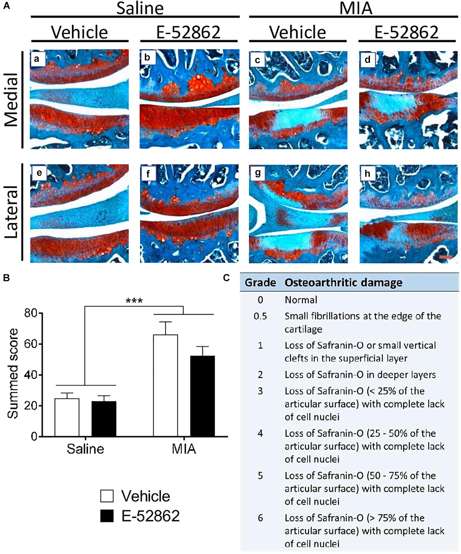
Monosodium iodoacetate is a chondrocyte glycolytic inhibitor which produces chondrocyte death and damage in the entire joint space. We determined the level of cartilage degeneration through proteoglycan staining 29 days after the intra-knee injection (Figure 2A). MIA injected mice had a clear increase on the OARSI score when compared to saline mice (p < 0.001; Figure 2B), and no significant effect of the E-52862 treatment (20 mg/kg, twice daily during 25 days) was found. Therefore, joint damage was not significantly prevented by the blockade of σ1R.
Histological knee alterations in mice injected with MIA were not prevented by the chronic treatment with E-52862.
Ipsilateral knees of saline and MIA mice were obtained 29 days after intra-articular injection in mice receiving vehicle or E-52862 treatment. (A) Medial and lateral sides of the joints are represented, showing the femur condyle (above) and the tibial plateau (below). (B) The injection of MIA produced cartilage degeneration revealed by an increased OARSI score. Treatment with E-52862 (20 mg/kg, twice daily during 25 days) did not prevent the joint damage. (C) The semiquantitative scoring system for joint histopathology. The scores for each image are (first value represents femur condyle and second value represents tibial plateau): (a) 1, 0.5; (b) 0.5, 0; (c) 2, 5; (d) 2, 5; (e) 2, 0.5; (f) 0, 0.5; (g) 3, 6; (h) 2, 3. Data are expressed as the mean ± SEM (n = 5–7 animals per group). Scale bar: 100 μm. ∗∗∗p < 0.001 for saline vs. MIA (2-way ANOVA). MIA, monosodium iodoacetate; SEM, standard error of the mean.
Acute and Chronic Blockade of σ1R Avoid Osteoarthritis-Induced Cognitive Impairment
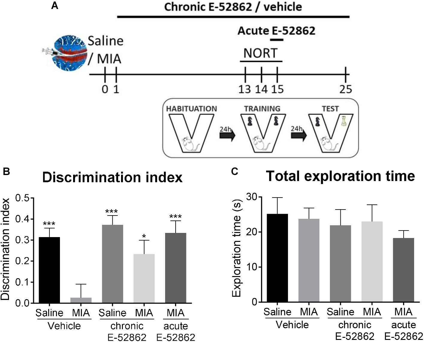
Chronic pain is often accompanied by memory dysfunction. Therefore, we analyzed the effect of chronic treatment with E-52862 (20 mg/kg, twice daily during 25 days) over recognition memory in the osteoarthritis model (Figure 3A). The novel object recognition task performed 15 days after MIA/saline injection showed a significant decrease on the discrimination index of MIA-injected mice treated with vehicle (p < 0.001 vs. saline). This cognitive impairment was avoided after the chronic treatment with E-52862 (p < 0.05 vs. MIA vehicle; Figure 3B). Interestingly, MIA-injected mice receiving a single acute dose of the σ1R antagonist (20 mg/kg) 30 min before the test also showed an improvement on the discrimination index (p < 0.001 vs. MIA vehicle) (Figure 3B). All groups of mice showed similar total exploration times, suggesting normal locomotor activity in this paradigm regardless of the surgery or the treatments (Figure 3C). Therefore, the impairment of recognition memory caused by chronic osteoarthritis pain was improved after chronic or acute blockade of σ1R.
Acute and chronic treatments with E-52862 improved the cognitive deficits induced by MIA injection.
(A) Saline or MIA-injected mice treated with vehicle or E-52862 (20 mg/kg, twice daily during 25 days) were evaluated for recognition memory 15 days after the intra-knee injection in the novel object recognition test (NORT). (B) Mice with osteoarthritis pain treated with vehicle showed decreased discrimination index indicating a memory impairment. Acute and chronic treatment with E-52862 reversed the cognitive deficits induced by MIA. (C) Animals revealed similar total exploration times regardless of the surgery or the treatment. Data are expressed as mean ± SEM (n = 6–8 animals per group). ∗p < 0.05, ∗∗∗p < 0.001 vs. MIA-vehicle (1-way ANOVA followed by Fisher least significant difference test). MIA, monosodium iodoacetate; SEM, standard error of the mean.
E-52862 Decreases Depressive-Like Behaviour Associated to Osteoarthritis Pain
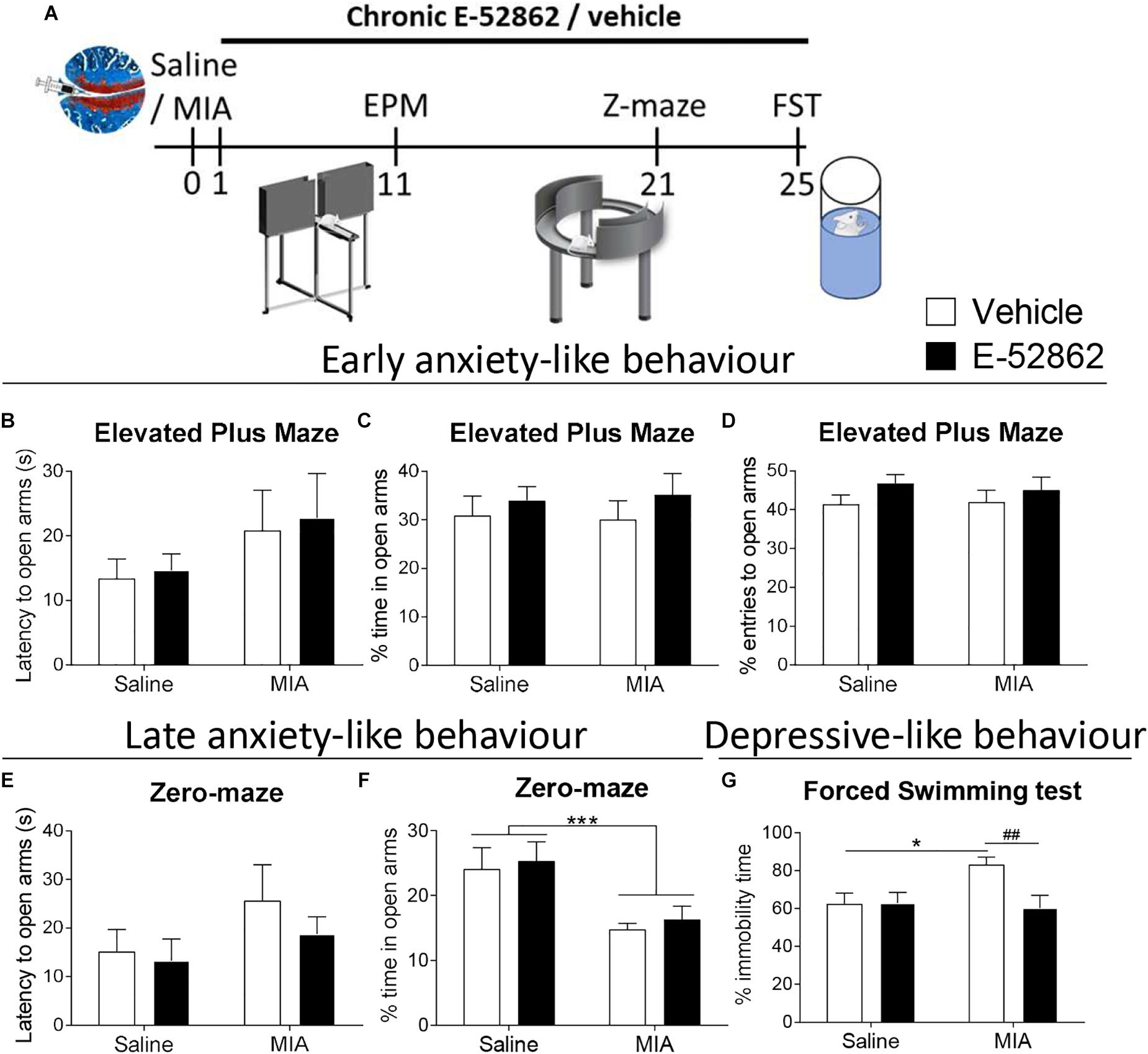
Anxiety and depressive-like behavior were assessed to determine whether E-52862 (20 mg/kg, twice daily during 25 days) could modulate emotional-like states associated to osteoarthritis pain (Figure 4A). It has been proposed that the initial stages of osteoarthritis pain are associated with inflammatory processes, whereas later stages involve neuropathic components, which may differentially affect the emotional manifestations. Thus, early and late anxiety-like behavior was assessed in our model. Early anxiety was evaluated 11 days after intra-knee injection in the elevated plus maze. No differences were found between saline- and MIA-injected mice in the latency to entry to the open arms, and the percentage of time and entries to the open arms, regardless of the treatment received (Figures 4B–D). On the other hand, despite the latency to the open quadrants of the zero-maze was not altered (Figure 4E), mice with osteoarthritis pain showed late anxiety-like behavior reflected in a significant decrease of the time spent in the open arms of the zero-maze (p < 0.001 vs. saline), also regardless of the treatment. Thus, E-52862 did not normalize the anxiogenic-like responses induced by MIA (Figure 4F). Depressive-like behavior was analyzed in the forced swimming test 25 days after the intra-articular injection. In this paradigm, mice with osteoarthritis pain receiving vehicle showed a significant increase on immobility time (p < 0.05 vs. saline; Figure 4G). Chronic E-52862 administration prevented such an increase in despair-like behavior (p < 0.01 vs. MIA vehicle; Figure 4G). Therefore, anxiety-like behavior was not sensitive to σ1R antagonism, whereas MIA-induced depressive-like behavior was prevented after E-52862 treatment.
E-52862 treatment reduced depressive-like behavior, but not anxiety-like responses associated with chronic osteoarthritis pain.
(A) Emotional manifestations of osteoarthritis pain were assessed in saline- or MIA-injected mice after repeated administration with vehicle or E-52862 (20 mg/kg, twice daily during 25 days) to saline or MIA-injected mice. Anxiety-like behavior was evaluated on day 11 after the intra-knee injection with the elevated plus maze (EPM), and at day 21 in the zero-maze (Z-maze), while depressive-like behavior was determined in the forced-swimming test (FST) on day 25. The latency to enter in the open arms (B), and the percentage of time (C) and entries (D) to the open arms of the EPM showed no significant differences between groups. At day 21, no significant differences were observed in the latency to the open quadrants of the zero-maze (E), whereas mice injected with MIA and treated with vehicle spent less time in the open parts (F). This increase on late anxiety-like behavior was not modified by E-52862 treatment. (G) Mice with osteoarthritis pain receiving vehicle showed increased immobility time, which was reversed by E-52862 treatment. Data are expressed as mean ± SEM (n = 6–8 animals per group). For (D): ∗∗∗p < 0.001 for saline vs. MIA (two-way ANOVA). For (E): ∗p < 0.05 for saline – vehicle vs. MIA – vehicle, ##p < 0.01 for MIA – vehicle vs. MIA – E-52862 (two-way ANOVA). MIA, monosodium iodoacetate; SEM, standard error of the mean.
E-52862 Modulates Microglial Expression in the Medial Prefrontal Cortex
A possible central role of σ1R modulating microglial activity was assessed through quantification of the density of microglial cells and the perimeter of the somas in the prelimbic and infralimbic areas of the medial prefrontal cortex (Figure 5). The analysis of the cellular density showed a significant increase on the total number of microglial cells in the prelimbic and the infralimbic areas of mice with osteoarthritis pain receiving vehicle (p < 0.001 vs. saline) (Figures 5A,C,D,F). Repeated administration of the σ1R antagonist (20 mg/kg, twice daily during 25 days) significantly reduced the microglial density in both cortical areas (p < 0.01 vs. MIA vehicle; Figures 5A,C,D,F). MIA-injected mice had an increase of the perimeter of microglial cells in the infralimbic (p < 0.05; Figure 5E), but not in the prelimbic area (Figure 5B) when compared to saline-injected mice. This increase was not significantly affected by the treatment with E-52862. Therefore, E-52862 modulated the density of microglial cells in the medial prefrontal cortices without affecting microglia activation.
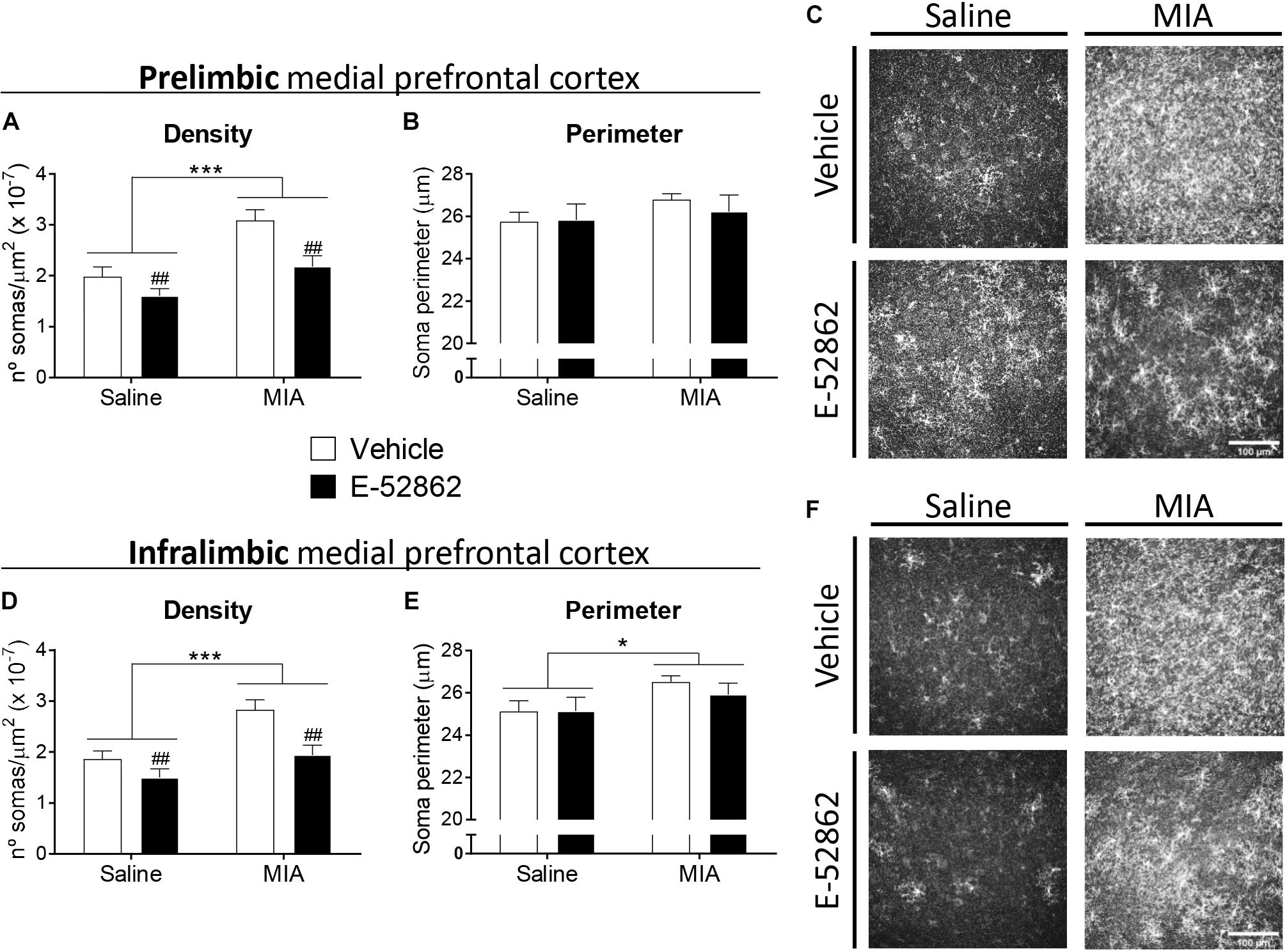
E-52862 decreased pain-induced microgliosis in the medial prefrontal cortex.
Microglial cells were detected in the prelimbic and infralimbic areas of the medial prefrontal cortex (mPFC) of saline and MIA injected mice after the repeated treatment with vehicle or the σ1R antagonist E-52862 (20 mg/kg, twice daily during 25 days). Mice with osteoarthritis pain treated with vehicle showed an increased density of microglial cells in the prelimbic (A) and the infralimbic areas (D). This microgliosis was normalized by the treatment with E-52862 (A,D). MIA-injected mice revealed larger perimeters of the soma of microglial cells in the infralimbic (E), but not in the prelimbic area (B). Treatment with E-52862 did not modify this alteration (E). (C,F)Representative images of all groups are shown. Data are expressed as the mean ± SEM (n = 7 animals per group). Scale bar: 100 μm. ∗p < 0.05, ∗∗∗p < 0.001 for saline vs. MIA, ##p < 0.01 for vehicle vs. E-52862 (two-way ANOVA). MIA, monosodium iodoacetate; SEM, standard error of the mean.
Discussion
The present study reveals the involvement of the σ1R in the nociceptive, emotional and cognitive alterations associated with osteoarthritis pain in mice. Mechanical allodynia and gait impairments induced by MIA injection were partly prevented by chronic administration of the σ1R antagonist E-52862. This treatment also inhibited the cognitive deficits and depressive-like behavior of mice with osteoarthritis pain, although anxiogenic-like responses were not modified. Modulation of the pain-induced behavioral alterations by E-52862 was not due to an inhibition of joint damage produced by MIA, and there was a concomitant decrease on MIA-induced microgliosis in the medial prefrontal cortex.
σ1R is highly expressed in key areas for pain control (Alonso et al., 2000; Bangaru et al., 2013). Behavioral studies have shown analgesic efficacy of the σ1R antagonist E-52862 in acute (Romero et al., 2012; Gris et al., 2014; Tejada et al., 2014) and chronic (Gris et al., 2014) models of inflammatory pain, and in neuropathic pain models induced by partial sciatic nerve ligation (Romero et al., 2012), chemotherapy (Nieto et al., 2012), or streptozotocin-induced diabetes (Gris et al., 2016). However, the role of σ1R has not been previously assessed in models of osteoarthritis pain, one of the most prevalent and disabling chronic pain conditions. We showed that E-52862 inhibited both mechanical hypersensitivity and gait alterations in the MIA model of osteoarthritis pain. Gait alterations could be associated to structural modifications of the joint or to the increased mechanical sensitivity (Boettger et al., 2009). Previous studies using the antigen-induced arthritis model in rats suggested that specific gait parameters, such as the angle between the paws, were exclusively influenced by the structural damage of the joint as indicated by its correlation with cartilage destruction (Boettger et al., 2009). However, other parameters, such as the paw print area, represent good measures of pain (Boettger et al., 2009). The correlation with mechanical allodynia would be in agreement with previous work showing that nerve-injured rats with decreased mechanical thresholds to punctate stimulation had also altered walking patterns (Vrinten and Hamers, 2003). In the same line, the MIA model of osteoarthritis pain in rodents showed that celecoxib and morphine reduced mechanical allodynia and gait abnormalities (Ferland et al., 2011; Ferreira-Gomes et al., 2012), suggesting that both parameters are associated in this chronic pain model. Such correlation has also been described in higher order mammals with osteoarthritis pain (Haussler et al., 2007; Frost-Christensen et al., 2008; Moreau et al., 2011; Cake et al., 2013). Thus, the reduction of the paw print area and the maximal contact area parameters observed in our study in osteoarthritic mice were probably a consequence of an unwillingness of the animal to bear weight on the injured limb, while the normalization of such parameters after E-52862 treatment might be related to reduced pain perception. In agreement, the effect of E-52862 on the walking patterns of mice with osteoarthritis was not accompanied by a normalization of the structural alterations observed in the histological assessments. This absence of effect on cartilage damage is in agreement with the low expression levels of σ1R in chondrocytes and bone marrow when compared to its expression in the peripheral and central nervous system1,2. The relief of mechanical hypersensitivity and pain-associated comorbidities after the treatment with E-52862 coexisted with the cartilage degradation, in agreement with the widely recognized fact that the presence and severity of joint pain poorly correlates with structural joint damage in osteoarthritis patients (Lawrence et al., 1966; Dieppe, 2004). Thus, the pain-relieving effects of the σ1R antagonist probably rely on a modulatory role on the nervous system and are independent of the site of the primary lesion.
We observed a cognitive deficit associated to osteoarthritis induced by MIA, which was significantly reduced by the repeated administration of E-52862. Our result suggests that the blockade of σ1R plays a protective role in this long-term memory impairment produced by chronic pain. Previous studies also showed impaired memory function in other chronic pain models (Zhao et al., 2006; Kodama et al., 2011) and specifically during MIA-induced joint pain (La Porta et al., 2015; Negrete et al., 2017). Selective σ1R ligands failed to modify learning, consolidation or retention phases of the mnemonic process when administered to naïve animals (Hashimoto et al., 2007; Antonini et al., 2011), but σ1R activation reduced cognitive deficits associated with schizophrenia (Hashimoto et al., 2007), Alzheimer disease (Maurice et al., 1998; Antonini et al., 2011) or scopolamine treatment (Hiramatsu et al., 2002). In contrast, we observed that σ1R blockade reversed the memory impairment induced after MIA injection. The overlap between the neuroanatomical substrates implicated in both pain control and cognitive functions provides information about the development of memory deficits in patients with chronic pain (Moriarty et al., 2011). However, the precise causal mechanisms underlying the pain-related cognitive impairment are still unclear, and the role of the σ1R on this specific type of memory deficits has not been studied. Our data suggest that σ1R antagonists are efficient improving cognitive functions under a chronic pain state.
We obtained increased anxiety-like responses after the intra-knee injection of MIA, as previously reported in other murine models of inflammatory (Schellinck et al., 2003; Chen et al., 2013) and neuropathic pain (Benbouzid et al., 2008; Matsuzawa-Yanagida et al., 2008; La Porta et al., 2016). Anxiety-like behavior was present 3 weeks after MIA, but not at earlier time points (11 days). Previous studies suggested that persistent pain may trigger alterations in brain areas involved in affective responses, which over time may lead to emotional comorbidities including anxiety and depressive-like behavior (Narita et al., 2006; Suzuki et al., 2007; Seminowicz et al., 2009; Sellmeijer et al., 2018). In agreement, 25 days after the intra-knee injection of MIA depressive-like responses were observed in animals with osteoarthritis pain, as in previous studies investigating inflammatory and neuropathic pain (Hasnie et al., 2007; Suzuki et al., 2007; Norman et al., 2010; Negrete et al., 2017). Depressive-like responses were abolished after chronic administration of E-52862, although anxiety-like behavior was not modified with this σ1R antagonist. These results are in line with previous works studying affective behavior in σ1R knockout mice. In these studies, σ1R knockouts exhibited increased immobility in the forced swimming test, but normal anxiety-like behavior (Sabino et al., 2009), suggesting distinct roles of the receptor modulating depressive and anxiety responses. Common neuroplastic changes associated with chronic pain and emotional disorders were proposed as important routes for the onset and reciprocal aggravation of both pathologies (Sheng et al., 2017). Consequently, analgesic drugs such as opioids (Mague et al., 2003; Tenore, 2008) or benzodiazepines (Vollenweider et al., 2011) have been proposed as a treatment for chronic pain-induced depression, and antidepressants like selective serotonin reuptake inhibitors (SSRIs) (Tasmuth et al., 2002; Gebhardt et al., 2016) or tricyclic antidepressants (Rowbotham et al., 2005; Kopsky and Keppel Hesselink, 2012) exhibited antinociceptive effects under chronic pain conditions. The interest of σ1R ligands for the treatment of depressive states raised from the observation that several antidepressants had moderate to high affinity for σ1R sites (Schmidt et al., 1989; Itzhak et al., 1991; Narita et al., 1996). While some SSRIs such as fluvoxamine or venlafaxine have shown agonism for σ1R, others like sertraline may act as antagonists (Ishima et al., 2014). Moreover, the antidepressant efficacy of σ1R ligands may depend on the affective status of the animal, since the selective σ1R agonist PRE-084 reduced depressive-like behavior in adrenalectomized mice but lacked effect in naïve animals (Urani et al., 2001).
We observed an increased microgliosis in the medial prefrontal cortex produced by the injection of MIA. This result agrees with a previous study showing increases of microglial density in the infralimbic cortex of nerve-injured rats (Chu Sin Chung et al., 2017; Xu et al., 2017). Other brain areas such as the amygdala, periaqueductal gray (PAG) or hippocampus, have also shown increased gliosis during chronic pain conditions (Humo et al., 2019). Interestingly, a recent study on neuropathic pain showed enhanced expression of microglial markers in the prefrontal cortex accompanied by depressive-like behavior. Chronic minocycline attenuated both microglial activation and depressive-like responses (Xu et al., 2017). Previous studies have shown that the σ1R antagonist BD1047 attenuated microglial activation in the spinal cord in a model of bone cancer pain (Zhu et al., 2015), but the effect of σ1R on supraspinal microglia has not been assessed in chronic pain models. Our data show that σ1R antagonist E-52862 significantly reduced the density of microglia in medial prefrontal cortices of mice with osteoarthritis pain. This effect was not accompanied by a reduction of anxiety-like behavior, suggesting that this affective disturbance is not directly related to cortical microgliosis. However, these anatomical changes correlated with the cognitive performance and the depressive-like behavior, pointing toward an involvement of cortical microglia on both pain comorbidities. Therefore, σ1R-regulated cortical microgliosis might be crucial for the manifestation of cognitive and emotional alterations often present in chronic pain conditions. Indeed, antidepressant drugs such as SSRIs also have activity modulating microgliosis and reducing microglial production of tumor necrosis factor α and nitric oxide (Chung et al., 2011; Tynan et al., 2012). It is well known that σ1R modulates several signal transduction pathways, including the production of ATP, reactive oxygen species or mitogen-activated protein kinases (MAPK) (Zamanillo et al., 2013; Hayashi, 2015; Zhao et al., 2017). All these molecules have been identified as effective signals for microglial migration and activation (Biber et al., 2007; Fan et al., 2017), suggesting an indirect modulatory role of σ1R. In agreement, σ1R activation by methamphetamine induces a microgliosis that involves generation of reactive oxygen species and activation of the MAPK pathway (Chao et al., 2017).
The present study reveals that E-52862 alleviates the nociceptive, cognitive and emotional manifestations associated to chronic osteoarthritis pain. We provide evidence showing that the effect of σ1R over these manifestations of chronic pain is not associated to local changes in articular damage but is accompanied by modulation of microglial activity in the medial prefrontal cortex. Our data highlight the blockade of σ1R as an interesting pharmacological strategy for the simultaneous management of multiple aspects of chronic osteoarthritis pain.