
Soluble α-Synuclein Is a Novel Modulator of Alzheimer’s Disease Pathophysiology

By Megan E. Larson, Mathew A. Sherman, Susan Greimel, Michael Kuskowski, Julie A. Schneider, David A. Bennett, and Sylvain E. Lesné
Excerpt from the article published in Journal of Neuroscience 25 July 2012, 32 (30) 10253-10266; Corrected on May 26, 2022 DOI: https://doi.org/10.1523/JNEUROSCI.0581-12.2012
Editor’s Highlights
- Neurodegenerative diseases share the common feature of the misfolding and aggregation of proteins that are normally soluble under physiological conditions.
- In Alzheimer’s disease (AD), the prototypical neuropathological lesions include the presence of extracellular (EC) amyloid/senile plaques composed of amyloid-β (Aβ) peptide fibrils and intraneuronal formation of hyperphosphorylated tau aggregates called neurofibrillary tangles.
- However, 30–40% of AD cases present with additional signs of proteinopathies, including intracellular (IC) inclusions composed of α-synuclein (αSyn) known as Lewy bodies (LBs) and Lewy neurites (LNs)
- αSyn has been implicated in the pathogenesis of Parkinson’s disease, the Lewy body variant of AD, Lewy body dementia, and some forms of prion diseases.
- AD may not be a disease of two misfolded proteins but instead might result from at least a three-pronged attack on neurons by soluble Aβ, tau and αSyn.
- Soluble Aβ species alters soluble tau function and chemistry, thereby allowing soluble αSyn to accumulate.
- This deleterious elevation of αSyn then leads to a selective decrease in certain presynaptic vesicle proteins and a dissociation of the protein composition of these vesicles, impairing neurotransmitter release at synapses already under siege by soluble Aβ oligomers and abnormal tau species at postsynaptic sites.
Abstract
Recent evidence has emphasized soluble species of amyloid-β (Aβ) and tau as pathogenic effectors in Alzheimer’s disease (AD). Despite the fact that Aβ, tau, and α-synuclein (αSyn) can promote each other’s aggregation, the potential contribution of soluble αSyn to AD pathogenesis is unknown. Here, we found an approximate twofold increase over controls in soluble αSyn levels in AD brains in the absence of Lewy body cytopathology. Importantly, soluble αSyn levels were a quantitatively stronger correlate of cognitive impairment than soluble Aβ and tau levels. To examine a putative role for αSyn in modulating cognitive function, we used the Barnes circular maze to assess spatial reference memory in transgenic mice overexpressing human wild-type αSyn. The results revealed that an approximate threefold elevation of αSyn in vivo induced memory deficits similar to those observed in AD mouse models. The neurobiological changes associated with this elevation of soluble αSyn included decreases in selected synaptic vesicle proteins and an alteration of the protein composition of synaptic vesicles. Finally, a synergism between Aβ/APP and human tau seems to be responsible for the abnormal elevation of soluble αSyn in transgenic mice. Altogether, our data reveal an unexpected role for soluble, intraneuronal αSyn in AD pathophysiology.
Introduction
Neurodegenerative diseases such as Alzheimer’s, Parkinson’s, Huntington’s, and frontotemporal dementia share the common feature of the misfolding and aggregation of proteins that are normally soluble under physiological conditions. In the case of Alzheimer’s disease (AD), the prototypical neuropathological lesions include the presence of extracellular (EC) amyloid/senile plaques composed of amyloid-β (Aβ) peptide fibrils and intraneuronal formation of hyperphosphorylated tau aggregates called neurofibrillary tangles. However, 30–40% of AD cases present with additional signs of proteinopathies, including intracellular (IC) inclusions composed of α-synuclein (αSyn) known as Lewy bodies (LBs) and Lewy neurites (LNs) (Hamilton, 2000; Trojanowski, 2002). The presence of these additional lesions does not appear to be innocuous, as subjects presenting with the LB variant of AD generally display a more rapid rate of cognitive decline than subjects with AD alone (Olichney et al., 1998). Studies using experimental models have proposed that Aβ, tau, and αSyn may have synergistic adverse effects (Giasson et al., 2003a; Lee et al., 2004). Clinton et al. (2010) recently reported that overexpression of mutant forms of these proteins in a transgenic mouse model may promote the accumulation and aggregation of each other and accelerate cognitive dysfunction in humans.
Over the past decade, accumulating evidence suggests that soluble, nonfibrillar Aβ (McLean et al., 1999; Walsh et al., 2002; Kayed et al., 2003; Cleary et al., 2005; Lesné et al., 2006; Shankar et al., 2008) and tau (Ramsden et al., 2005; Santacruz et al., 2005; Roberson et al., 2007) assemblies may be the most neurotoxic species. To our knowledge, a putative role for soluble αSyn in AD has not been examined to date. However, several reports have shown that overexpression of wild-type αSyn (αSynWT) in vivo and in vitro leads to alterations of neuronal physiology. In an initial study, a threefold increase in human αSynWT in mouse brain led to the reduction of neurotransmitter release by inhibiting synaptic vesicle recycling (Nemani et al., 2010). Moreover, these transgenic animals displayed a selective decrease in specific synaptic vesicle proteins associated with the lowered presynaptic release (Nemani et al., 2010). Remarkably, both these phenomena occurred in the absence of any deposited, fibrillar αSyn (M. K. Lee et al., 2002). The second study identified an apparent alteration of the normal protein composition in synaptic vesicles when human αSynWT is transfected in neurons (Scott et al., 2010). The elevation of αSyn induced by the transfection did not lead to the formation of fibrillar aggregates, further validating the concept that dysregulation of soluble levels of αSyn can have crucial functional consequences for neuronal physiology.
Based on the aforementioned reports, we aimed to decipher whether abnormal alterations in soluble, nonfibrillar forms of αSyn occurred in AD and what functional and neurobiological consequences were associated with these potential changes.
…
Results
To determine the potential contribution of soluble αSyn in AD-associated memory decline in humans, we first measured its relative protein expression in the inferior temporal gyrus (ITG) of 84 elderly individuals from the ROS with no cognitive impairment (NCI), mild cognitive impairment (MCI), or AD. Neuropathological, cognitive, and epidemiological data are briefly summarized here (Table 1). The selection of the ITG for our analyses was guided by the following observations: (1) this region of the cerebral cortex shows reduced glucose utilization in AD and in asymptomatic individuals at risk genetically for AD (Small et al., 2000); (2) ITG gray matter thickness significantly predicts hippocampal volume loss in both amyloid-positive and hyperphosphorylated tau-positive individuals among MCI and AD individuals (Desikan et al., 2010); and (3) ITG amyloid loads and tangle density matched very well with average total brain amyloid burden (rho = 0.946; p < 0.0001) and tangle density (rho = 0.772; p < 0.0001).
Segregation of brain protein pools
To measure the various soluble pools of αSyn, we used an extraction protocol previously used to detect soluble Aβ oligomers in mouse brains (Lesné et al., 2006; Sherman and Lesné, 2011). Similar to the characterization of this method in mouse brain tissue, this procedure allows the study of different pools of a protein depending on its cellular compartmentalization (Lesné et al., 2006). Four protein fractions result from this method: the generated fractions are termed EC, IC, MB, and insoluble as they are, respectively, enriched in extracellular, intracellular, membrane-bound, and insoluble proteins. The first three fractions were subjected to ultracentrifugation at 100,000 × g for 1 h at 4°C, and supernatants containing soluble proteins were used for analyses. Human control proteins with well known compartmentalization were used to validate the method. Results are shown in Figure 1A, and proteins displayed a similar profile as observed previously. We then evaluated the segregation of several disease-related proteins, i.e., tau, APP, TAR DNA-binding protein-43 (TDP-43), the cellular prion protein PrPc, and αSyn (Fig. 1B). Consistent with existing knowledge about these proteins, we found that tau was mainly detected in the IC fraction (Fig. 1B, top inset) and TDP-43 was primarily present in the MB-enriched fraction (which also includes nuclear proteins as illustrated by NeuN compartmentalization in Fig. 1A), whereas it was barely detected in the IC-soluble extracts (Fig. 1B). The glycosylphosphatidylinositol-anchored cellular prion protein PrPc was only detected in the fraction enriched with membrane-associated proteins. The profile observed for αSyn was in agreement with our current knowledge of this protein (Fig. 1B, bottom inset): αSyn was primarily detected in EC and IC fractions. Consistent with previous observations (H. J. Lee et al., 2002), 10–15% of αSyn is membrane bound. Maybe more surprising is that EC levels of αSyn were just as high as IC levels, suggesting that a substantial portion of αSyn may be secreted in vivo (Lee et al., 2005). A recent report using microdialysis seems to confirm that αSyn is indeed secreted in the interstitial fluid of transgenic and human brain tissue (Emmanouilidou et al., 2011). In contrast, β-synuclein (βSyn) displayed a distinct segregation pattern compared with αSyn. βSyn was nearly completely localized in the IC fraction. βSyn was not detected in MB extracts, as expected, since βSyn lacks the hydrophobic core (VTGVTAVAQKT), allowing αSyn to interact with lipids.
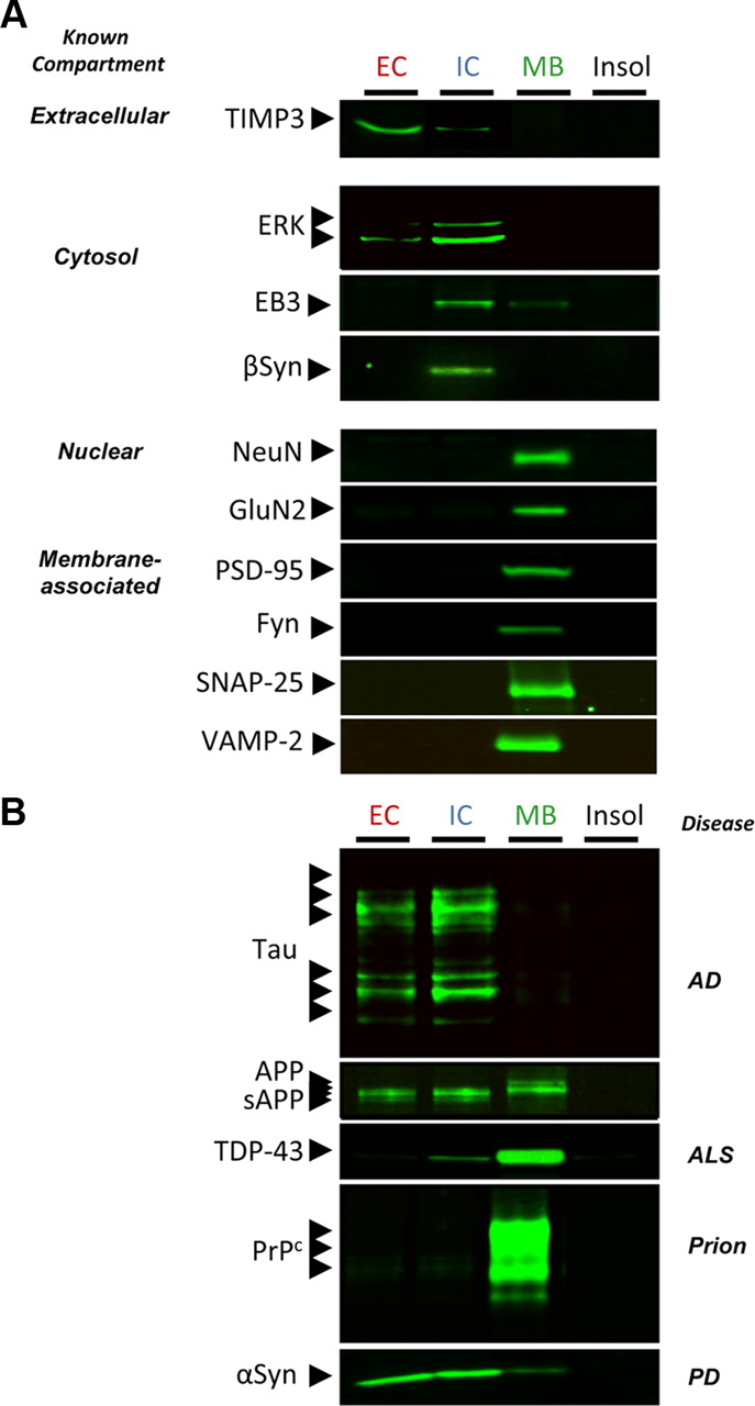
Characterization of the four-step protein extraction protocol using human brain tissue.
A, Selected brain tissue (0.5 mm3 of inferior temporal gyrus of a control brain) was subjected to serial extractions allowing the segregation of proteins based on their cellular compartmentalization. Examples of obtained segregation for human proteins with known compartmentalization are shown. B, Extraction profile for selected disease-related proteins. TIMP3, Tissue inhibitor of metalloproteinase-3; ERK, extracellular signal-regulated kinase; EB3, end-binding protein-3; NeuN, neuronal nuclei; GluN2, glutamate NMDA receptor subunit 2; PSD-95, postsynaptic density protein-95; Fyn, Fyn kinase; SNAP-25, synaptosomal-associated protein 25; VAMP-2, vesicle-associated membrane protein 2; sAPP/APP, soluble/full-length amyloid precursor protein; ALS, amyotrophic lateral sclerosis; PD, Parkinson’s disease; Insol, insoluble.
Altogether, these new results using human brain cerebral tissue and our past results using mouse brain tissue (Lesné et al., 2006, 2008) indicate that this biochemical method is suitable for measuring the respective soluble pools of brain proteins in human tissue.
Accumulation of soluble intracellular αSyn in AD
We first measured cerebral levels of the protein αSyn in our soluble fractions using specific commercially available antibodies, LB509, 4D6 (Figs. 1, 2), and BD42 (data not shown), after SDS-PAGE. However, whereas apparent SDS-resistant αSyn dimers (∼29 kDa) and pentamers (∼72 kDa) were observed in EC fractions, these αSyn oligomers were not readily detected in IC extracts, suggesting that oligomerization of αSyn might occur in considerable portion extracellularly in vivo. This observation is consistent with recent in vitro studies showing that αSyn oligomers can form extracellularly (Danzer et al., 2011). No other putative SDS-stable oligomeric forms of αSyn were readily detected by SDS-PAGE using LB509 (Fig. 2A) or BD42 (data not shown) or using antibodies targeting phosphorylated αSyn at serine 129 (pS129-αSyn) (data not shown). To ensure that these putative soluble αSyn oligomers were not artificially generated by SDS-PAGE, we separated brain protein EC extracts by gel filtration column and analyzed each eluted fraction using LB509 (Fig. 2B). Although it is believed that monomeric αSyn behaves as a disordered monomer leading to an elution of monomeric αSyn at ∼50 kDa in gel filtration experiments, a recent controversy has emerged as to what is the proper folding of αSyn under physiological conditions (Bartels et al., 2011; Fauvet et al., 2012). It is also unclear whether putative oligomeric forms of αSyn would follow a nonglobular elution profile, and we postulated that multimeric αSyn might behave as a globular protein similarly to Aβ molecules (Lesné et al., 2006). If this hypothesis were correct, we predicted that apparent SDS-resistant dimeric αSyn would elute within fractions corresponding to ∼28 kDa. Using either LB509 or 4D6 antibodies (data not shown), our results revealed that αSyn dimers did indeed elute at the expected theoretical size (28–29 kDa) for globular proteins in the absence of apparent monomeric αSyn. This finding indicated that putative SDS-resistant dimeric αSyn were present in our samples. For the reasons mentioned above, we determined the respective levels of three soluble αSyn molecules that we could reliably assess by SDS-PAGE: αSyn monomers in EC and IC fractions and αSyn dimers in the EC fraction (Fig. 2A,B). Of note is that because neuronal loss could potentially alter our measurements, we determined the cerebral expression levels of the neuron-specific nuclear protein NeuN and normalized all protein measurements to them. Under these conditions, we found that monomeric and dimeric αSyn-EC ITG levels were unchanged across clinical groups (Fig. 2C). Monomeric intracellular αSyn (1mer-IC) was remarkably increased in AD compared with the NCI and MCI groups (a 2.24- and a 1.69-fold elevation vs NCI and MCI, respectively; p = 0.0009).
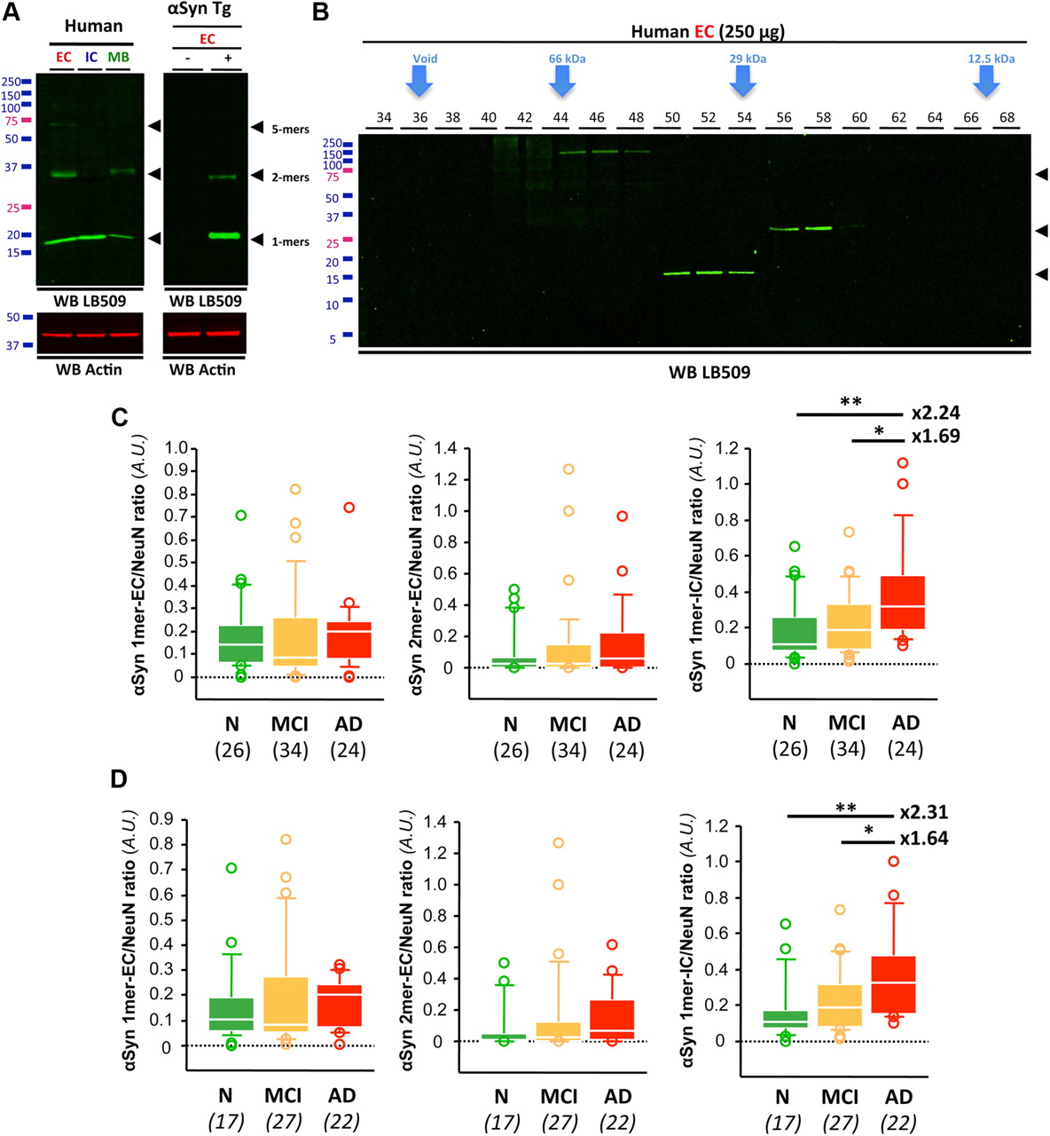
Increase in soluble αSyn in AD brain in the absence of Lewy bodies/neurites.
A, Western blot (WB) analyses of soluble αSyn species in EC-, IC-, or MB-enriched fractions using LB509. Similar results were obtained with 4D6 or Syn1 (not shown). Both transgenic and wild-type littermates from line G2.3-A53T (αSyn Tg) used were 10 months of age. B, Gel filtration combined with SDS-PAGE confirmed the presence of SDS-resistant αSyn soluble assemblies. C, Quantification of monomeric and oligomeric αSyn species in the inferior temporal cortex of subjects with NCI, MCI, or AD. Whereas monomeric and dimeric αSyn-EC remained unchanged across groups, monomeric αSyn-IC levels were significantly higher in AD than in MCI and NCI brain tissues. D, In total absence of LB pathology, αSyn-IC monomers were increased by more than twofold in AD compared with controls. Italicized numbers in parentheses indicate group sizes. NCI is shown in green, MCI is shown in orange, and AD in indicated by red boxes. In box plots of all figures, the bar inside the box indicates the median; the upper and lower limits of boxes represent the 75th and 25th percentiles, respectively; and bars flanking the box represent the 95th and 5th percentiles. *p < 0.05; **p < 0.01. N, no cognitive impairment.
Since αSyn is known to form inclusions in 30–40% of AD cases, we looked for such intracellular aggregates in our study participants. Inclusions were found in brain regions other than the one studied here (ITG) in 18 brains. To determine whether the observed increase in soluble monomeric αSyn-IC was a reflection of LB/LN pathology, we repeated our analyses excluding all of these specimens (Fig. 2D, the modified numbers of cases remaining after exclusion are indicated in italics). In the complete absence of immunohistochemically detectable αSyn pathology, the AD group still had a more than twofold increase in αSyn-IC monomers (2.31-fold elevation vs NCI; p < 0.0001), suggesting that this accumulation is independent of LB/LN formation. Similarly, we measured αSyn levels in the membrane-associated protein fractions and observed no changes across groups (data not shown).
To explore whether changes in gene expression were responsible for this elevation, SNCA mRNA levels were measured by reverse transcriptase-qPCR (rt-qPCR). Despite a lower number of tissue specimens available for transcriptional analyses, relative SNCA mRNA levels were increased by 1.67-fold in AD compared with age-matched controls (Fig. 3A).
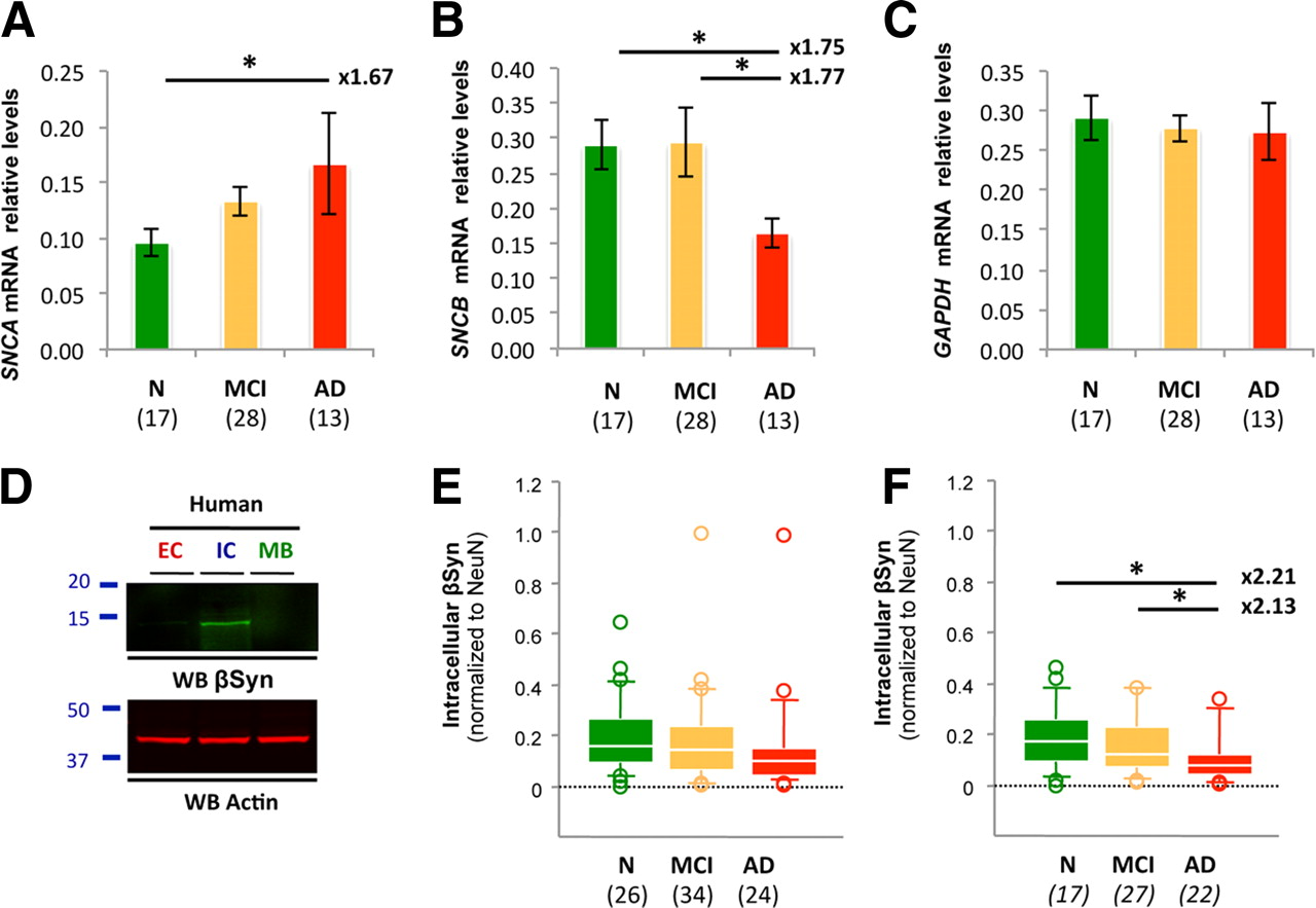
SNCA and SNCB gene expression in the ROS cohort. A–C, SNCA
(A), SNCB (B), and GAPDH (C) mRNA levels were measured by rt-qPCR and normalized to the housekeeping gene β-actin. GAPDH was used as the internal control. Quantification revealed an ∼1.7-fold increase in SNCA gene expression in AD compared with age-matched controls, consistent with the approximate twofold elevation of soluble αSyn observed at the protein level. SNCB mRNA levels were lowered by ∼1.8-fold. D, Western blot (WB) analyses illustrating the biochemical segregation of soluble βSyn in EC-, IC-, or MB-enriched fractions. E, Quantification of βSyn in the inferior temporal cortex of subjects with NCI, MCI, or AD. No statistical changes were observed. F, In the absence of LB pathology, βSyn levels were reduced by approximately twofold in AD compared with controls. Values represent mean ± SD (ANOVA followed by Fisher’s PLSD test, *p < 0.05). Italicized numbers in parentheses indicate group sizes. NCI is shown in green, MCI is shown in orange, and AD in indicated by red boxes. In box plots of all figures, the bar inside the box indicates the median; the upper and lower limits of boxes represent the 75th and 25th percentiles, respectively; and bars flanking the box represent the 95th and 5th percentiles. *p < 0.05; **p < 0.01. N, no cognitive impairment.
Because the β isoform of synuclein (βSyn) is believed to act as a negative regulator of αSyn (Hashimoto et al., 2001; Snyder et al., 2005), we measured SNCB mRNA and protein expression in our cohort (Fig. 3B–F). At both levels, a 2.2- and a 1.7-fold decrease were, respectively, observed in AD compared with age-matched nonimpaired subjects. These results suggest an imbalance of SNCA/SNCB gene expression in AD.
Soluble αSyn levels are linked to AD-associated cognitive impairment
To assess the potential impact of soluble αSyn in AD, we examined correlations between measures of cognitive function (episodic memory, semantic memory, working memory, perceptual speed, visuospatial ability, global cognition) and measures of αSyn protein in ITG (total, n = 84; Table 2, color map matrix). Soluble monomeric Aβ levels and soluble total tau levels were used as internal controls. Both were determined using the same technique used for αSyn to avoid intrinsic differences between detection methods. Of interest is that buffer-soluble Aβ levels measured by Western blotting closely matched levels observed by ELISA (rho = 0.646; p < 0.0001). As reported previously, soluble Aβ ITG levels were inversely correlated with multiple memory modalities, notably episodic and global cognition (Table 2, matrix, first column). Soluble total tau levels (using Tau5) seemed to reflect a more profound effect, as these levels were linked to two additional modalities (Table 2, matrix, second column). These results are in general agreement with our current understanding of AD pathophysiology (Bennett et al., 2004; Jack et al., 2010) and further validate the use of the ITG.
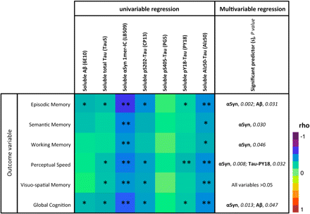
Summary of regression models for soluble neuropathological proteins in Alzheimer’s disease
All measures of soluble Aβ, tau, and αSyn were performed using the same technique (SDS-PAGE followed by Western blot) to avoid inherent differences between techniques. Models were performed using the entire cohort (n = 84). *Correlation is significant at the 0.05 level (two-tailed); **Correlation is significant at the 0.01 level (two-tailed). The significant predictor is in bold, the p value is in italics.
In contrast to the significant correlations of soluble Aβ or soluble tau molecule with just some cognitive measures, soluble αSyn (1mer-IC) in ITG was inversely correlated to all measures of cognitive function (Table 2, matrix, third column). Since hyperphosphorylation of tau is a key change in AD pathophysiology, we assessed the ITG levels of disease-relevant tau epitopes/conformations, i.e., pS202-Tau, pS405-Tau, pY18-Tau, and Alz50-Tau using CP13, PG5, PY18, and Alz50, respectively (kind gifts from Drs. Peter Davies, Albert Einstein College of Medicine, Bronx, NY, and Gloria Lee, University of Iowa, Iowa City, IA). Although pS405-Tau levels showed no apparent relationship to cognitive function, an early marker of tau pathological changes, pS202-Tau, displayed a similar regression profile to total soluble tau (Table 2, matrix, fourth column). Alz50-Tau levels were inversely correlated to all measures of cognitive function (Table 2, matrix, seventh column). Even though the profile observed for Alz50-Tau was similar to soluble αSyn, the relative strength of the correlations indicated by the rho value seemed stronger for αSyn compared with Alz50-Tau. This unexpected and surprising finding would suggest that soluble intracellular αSyn might be an important correlate of decreased cognitive function in AD.
Soluble αSyn as a good predictor of AD-related impairment
Recent genetic evidence indicates that Aβ, tau, and αSyn proteins potentiate each other’s aggregation (Clinton et al., 2010). However, it is not known whether this observation also applies to the soluble forms of these proteins. Here, we noticed that cortical levels (by quantitative Western blotting) of soluble forms of Aβ, tau, and αSyn were positively correlated to each other (Table 3), suggesting that the respective levels of these soluble proteins might influence each other. Surprisingly, soluble αSyn was strongly correlated to total Tau (rho = 0.527; p < 0.0001) and more modestly related to soluble Aβ (rho = 0.268; p = 0.0141) and pS202-Tau (rho = 0.228; p = 0.0376). Note that soluble tau, however, was correlated to all disease-relevant tau species.
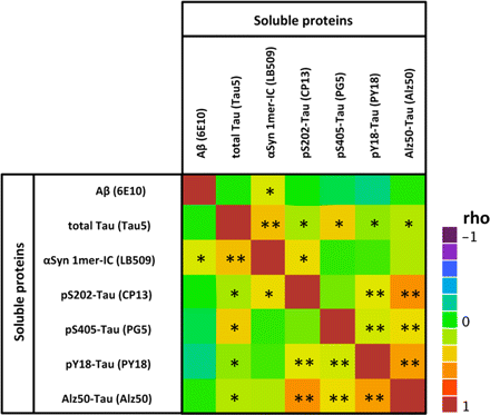
Color map on Spearman rho correlations for select soluble proteins in Alzheimer’s disease
All measures of soluble Aβ, tau, and αSyn were performed using the same technique (SDS-PAGE followed by Western blot). Models were performed using the entire cohort (n = 84). *Correlation is significant at the 0.05 level (two-tailed); **Correlation is significant at the 0.01 level (two-tailed).
Because of these intercorrelations, multivariate linear regression models were used to estimate the independent effects of specific protein variables (all log transformed) as predictors of cognitive measures (episodic memory, working memory, perceptual speed, visuospatial memory, and global cognition) (Table 2, far-right column). Soluble αSyn (1mer-IC) was a significant independent predictor (all p < 0.05) of scores on episodic memory, semantic memory, working memory, perceptual speed, and global cognition. For semantic and working memory outcomes, soluble αSyn was the only significant predictor variable in the multivariable models. Soluble Aβ monomers were a significant predictor for episodic memory (p = 0.031) and global cognition (p = 0.047). No variable was significant in the multivariable regression model for visuospatial memory, but soluble αSyn monomers approached significance as a predictor variable (p = 0.10). When it was the only predictor (in the univariable predictor model), it was statistically significant (p = 0.006). There was no evidence of multicollinearity problems in the multivariable models as assessed by the variance inflation factor (data not shown). Altogether, our findings indicate that increases in soluble αSyn monomers may contribute to the development of cognitive symptoms in AD.
Increased expression of αSyn causes memory impairment in transgenic mice
To address whether an elevation of soluble αSyn is sufficient to induce memory deficits in the absence of LB formation, we measured spatial reference memory in the Barnes circular maze (Fig. 4) using TgI2.2 mice, which overexpress human wild-type αSyn but do not form LBs (M. K. Lee et al., 2002). Accordingly, this line does not display apparent motor deficits (M. K. Lee et al., 2002). The well characterized J20 line, a common transgenic mouse model of AD (Mucke et al., 2000), was used as a positive control for spatial memory impairment. Both lines were compared with background strain-matched nontransgenic littermate mice. Whereas all three groups acquired the task equivalently (Fig. 4A,B), retention was greatly altered in Tg-I2.2 mice (Fig. 4C–E). In fact, memory performance was as compromised in this αSyn-overexpressing line than in the APP transgenic J20 mice. To rule out faster extinction in Tg-I2.2 mice compared with J20 and nontransgenic mice, we analyzed probe trial performance by segments of 60 s (Fig. 4F,G). Our data indicated that none of the three groups tested showed signs of extinction during the duration of the probe (180 s). In agreement with similar findings obtained in a different transgenic line (Magen et al., 2012), these results demonstrate that overexpression of soluble αSyn, in the absence of fibrillar pathology, is associated with memory deficits.
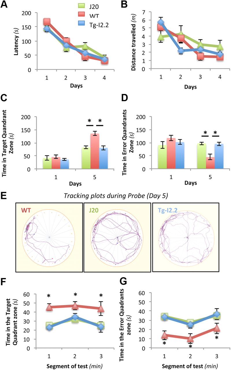
In the absence of pathology, overexpression of wild-type human αSyn causes memory deficits in the Barnes circular maze. Seven-month-old nontransgenic C57BL6, J20, and Tg-I2.2 mice (n = 4 mice per genotype) were trained in the Barnes circular maze for 4 d. A probe trial (escape platform removed) was conducted 24 h after the last training session. A, B, During acquisition of the task, latency (A) and distance traveled (B) were recorded. All three groups learned this task. Two-way repeated-measures ANOVA (RMANOVA) revealed a significant effect of training (F = 95.61; p = 0.0004) but no effect of transgene (F = 0.015; p = 0.9058) and no significant day × transgene interaction (F = 1.10; p = 0.3827). C–E, During the probe trial on day 5, wild-type mice, but not mice with hAPP or hαSyn, displayed a search bias for the target quadrant (C, D) and spent less time in nontarget quadrants (D, E). Two-way ANOVA of these data revealed a significant effect of hAPP (C, F(1,6) = 4.32, p = 0.038; E, F(1,6) = 4.68, p = 0.015) and hαSyn (C, F(1,6) = 14.34, p = 0.009; E, F(1,6) = 13.45, p = 0.010) and significant day × transgene interactions for both transgenic lines (C, FJ20(4,24) = 3.916, p = 0.013 and FI2.2(4,24) = 4.64, p = 0.041; E, FJ20(4,24) = 5.08, p = 0.004 and FI2.2(4,24) = 2.88, p = 0.044). F, G, Neither J20 nor TgI2.2 mice showed patterns of faster extinctions than nontransgenic animals during the 3 min of the probe trial, as indicated by the average time spent in the target quadrant or nontarget quadrants across 1 min intervals. Data represent mean ± SEM (n = 6 males per age per genotype; univariate RMANOVA test).
Elevation of soluble αSyn is associated with a selective reduced expression of synaptic vesicular proteins
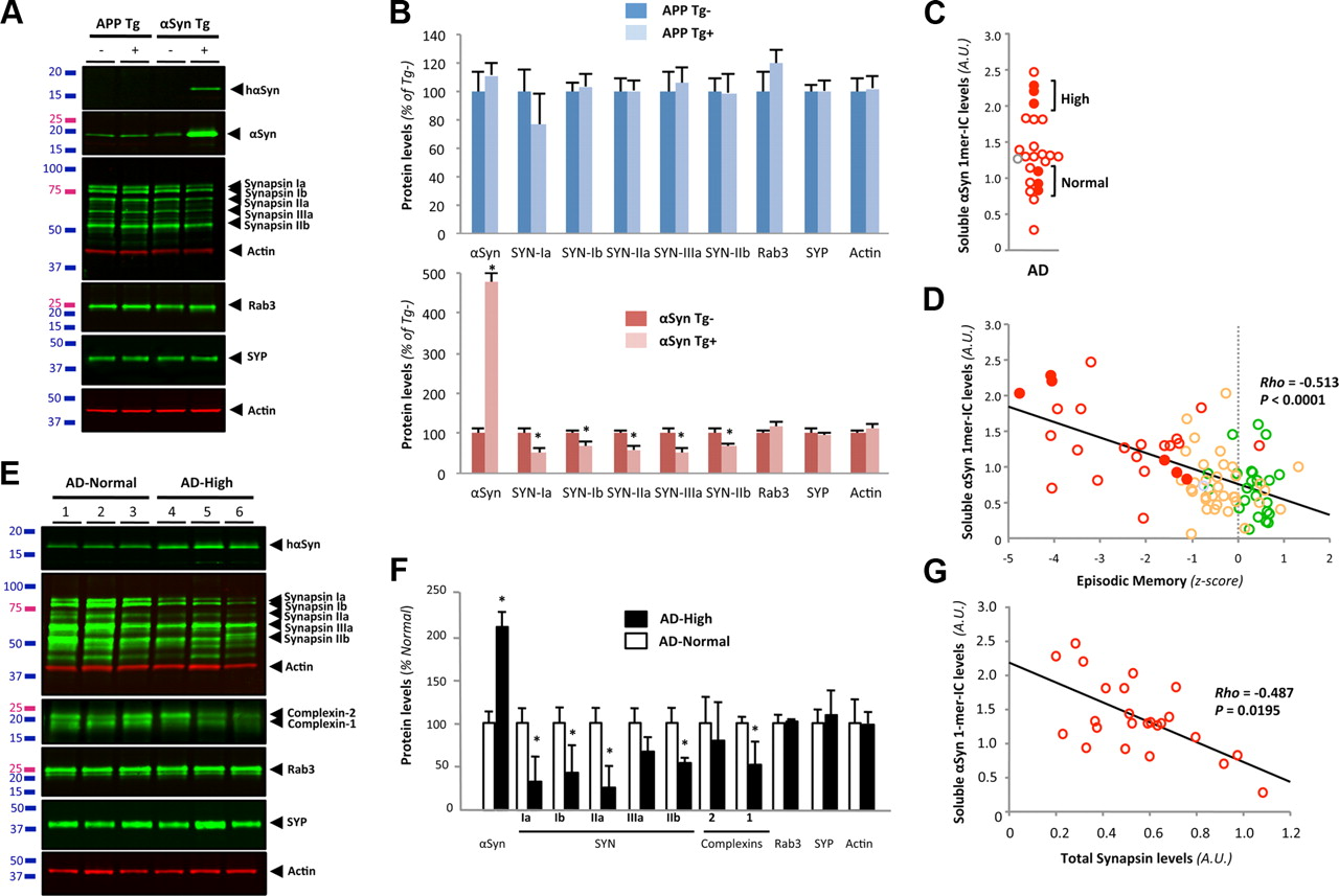
Since αSyn emerged as the strongest biological correlate of decreased cognition among the variables tested, we sought to identify the neurobiological changes underlying these relationships. A recent report demonstrated that a threefold increase in wild-type human αSyn (αSynWT) levels in the transgenic line I2-2, which does not lead to the formation of LBs/LNs (M. K. Lee et al., 2002), caused selective decreases in the expression of several presynaptic proteins controlling neurotransmitter release (Nemani et al., 2010). The downregulated proteins in this mouse line were synapsins (SYN) I/II and complexins 1/2, whereas levels of other synaptic vesicle proteins such as Rab3 and synaptophysin remained unchanged. We first compared the expression changes of the same presynaptic vesicular proteins in the most commonly used AD mouse model Tg2576 (Hsiao et al., 1996) and in the widely used Parkinson’s disease model αSynA53T TgG2-3 line (M. K. Lee et al., 2002) (Fig. 5A). In 17-month-old Tg2576 mice, none of the presynaptic vesicle proteins measured was altered compared with nontransgenic animals (Fig. 5A, lanes 1 and 2, B). Of note, we also found no changes at earlier ages, i.e., 1, 3, 6, 12, and 15 months, in this model (data not shown). In asymptomatic TgG2-3 mice devoid of LB/LN pathology, we found an approximate fivefold increase of αSyn expression over endogenous levels, in agreement with previously published results (M. K. Lee et al., 2002). Contrary to the Tg2576 APP mice, we noticed a marked reduction of synapsins (Fig. 5A, lanes 3 and 4, B) and complexins (data not shown) in the TgG2-3 αSyn mice. Neither Rab3 nor synaptophysin nor actin expression was changed. Having demonstrated that we could detect selective changes in synaptic vesicle protein expression induced by transgene-derived αSyn, we then asked whether the mean twofold elevation of intracellular monomeric αSyn levels seen in our AD group was sufficient to lead to similar abnormalities in human brains. Because our AD group contained an interindividual variability compatible with selecting two subgroups with relative αSyn soluble monomers levels differing by a factor of ∼2, we selected three brains representing the AD-High group (“high” levels of soluble αSyn) and three brains representing the AD-Normal group (“normal” αSyn levels) (Fig. 5C, filled circles; n = 3 for each group). When plotted against episodic memory performance (Fig. 5D), the AD-High subgroup had a worse mean z-score than the AD-Normal subgroup (−4.29 ± 0.39 vs −1.345 ± 0.23, respectively). We then analyzed the expression levels of synaptophysin, synapsins (Ia/b, IIa/b), complexins (−1/−2), Rab3, and the housekeeping gene product actin (Fig. 5E). In AD-High group, we saw significant decreases in levels of synapsins and complexin-1 but not synaptophysin, Rab3, and actin (Fig. 5E,F). These results mirror the alterations seen in the transgenic I2-2 mice overexpressing αSyn and having neurotransmitter release deficits (Nemani et al., 2010; Scott et al., 2010) in the absence of LB/LN formation. To further validate the apparent relationship between synapsin and αSyn expression in this small number of cases (n = 3 per group), we expanded our analyses to our entire AD cohort (n = 24). By quantitative Western blot, synapsin levels were inversely correlated to soluble αSyn monomer levels (Fig. 5G; rho = −0.487; p= 0.01).
A twofold increase in soluble αSyn is associated with a selective decrease in vesicular proteins in AD.
A, Western blot (WB) analysis of brain extracts from 17-month-old Tg2576 (APP Tg) and 10-month-old TgG2.3-A53T (αSyn Tg) brain tissue using infrared-conjugated secondary antibodies shows a selective reduction in synapsins in αSyn Tg. Rab3, synaptophysin, and actin levels were unchanged in both lines. B, Quantification of protein levels detected in both lines revealed specific decreases in synapsins in αSyn Tg mice (n = 3/group). C, Scatterplot of soluble αSyn levels in the ITG of subjects with AD. Measurements reflect the quantification of αSyn by WB after SDS-PAGE using the antibody LB509. Selected AD subjects were chosen to compose two groups (n = 3 per group) whose soluble αSyn ratio equaled 2. They are referred to as AD-High and AD-Normal and are indicated by filled red circles. D, Linear regression depicting the relationship between episodic memory and the levels of monomeric αSyn in the IC fraction measured by SDS-PAGE. Filled circles indicate specimens selected for analyses of vesicular presynaptic proteins (see Figs. 3 and 4). Note that the AD subjects with a twofold elevated αSyn are more impaired than AD subjects with lower αSyn expression. E, Quantitative WB analysis of brain extracts from AD brain tissue with normal (AD-Normal) or high (AD-High) (2-fold increase) αSyn-IC levels shows a reduction in synapsins and complexins but not Rab3 or synaptophysin. F, Quantification confirms the observed changes in B. Values represent mean ± SD (n = 3). G, Regression analyses between total synapsin protein expression and “monomeric” αSyn in all AD cases (n = 24) indicated a negative correlation (Spearman rho).
Dissociation between αSyn and synapsins in presynaptic vesicles in absence of LB
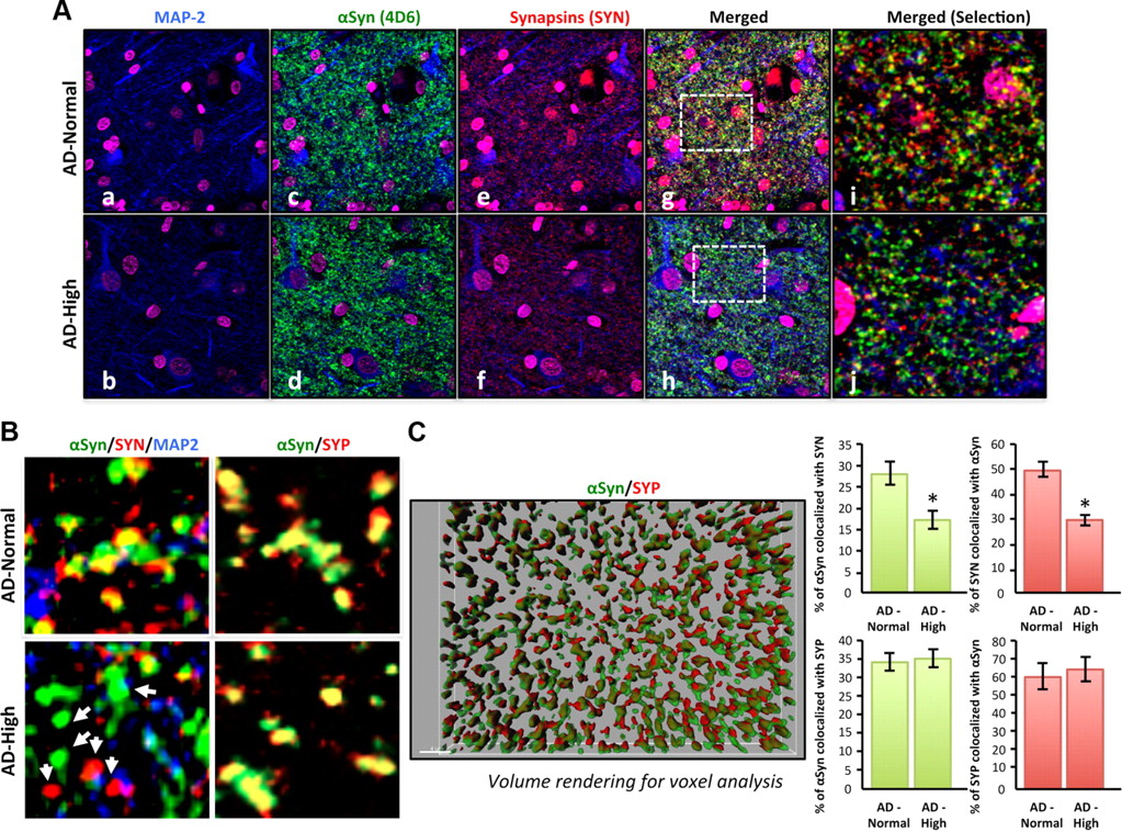
Both in brains of subjects with αSyn pathology [dementia with Lewy body (DLB)] and in primary hippocampal neurons derived from LB-forming PDGF-h-α-syn:GFP mice (Rockenstein et al., 2005), the elevation of neuronal αSyn levels is accompanied by enlarged presynaptic vesicles and dissociation of the colocalization pattern of αSyn with synapsins in synaptic boutons (Scott et al., 2010). On the basis of our results above, it seems that some pathological form(s) of αSyn can lead to a loss of certain synaptic vesicle proteins, which in turn induces functional synaptic deficits. It is, however, not clear whether soluble or insoluble αSyn is responsible for these abnormal alterations, since both disorders (human DLB and αSyn transgenic mice) display the presence of αSyn inclusions and/or phosphorylation at Serine 129 linked to fibrillar, insoluble αSyn aggregates. To test whether elevated levels of soluble αSyn are associated with changes in proteins normally expressed in presynaptic vesicles in the ITG in AD, we performed immunohistochemical colocalization for αSyn and synapsins in the same AD brains used for Figure 6, B and C. Representative laser-scanning confocal images are shown for the microtubule-associated protein MAP-2 (Fig. 6Aa,b, blue), αSyn (Fig. 6Ac,d, green), and synapsins (Fig. 6Ae,f, red). In agreement with our previous neuropathological assessments, we observed no LBs or LNs in the AD brains selected for the AD-Normal or AD-High subgroups (data not shown). Merged channels are shown at low (40×; Fig. 6Ag,h) and high (60×; Fig. 6Ai,j) magnification; yellow indicates colocalization between αSyn and synapsins in the same synaptic vesicles. Even in raw images, dissociation between these two proteins was obvious (Fig. 6Ag–j). To measure the imbalance in protein composition in synaptic vesicles between these subgroups, images were subjected to specific thresholds before software-operated quantification of the colocalized signals for αSyn and synapsins. Examples of such images are displayed in Figure 3B. Quantification confirmed a significant decrease (up to 32.8%) in vesicles immunoreactive for both αSyn and synapsins in the AD-High subgroup (Fig. 6B; Student’s t test, p < 0.0001). Importantly, none of the αSyn antibodies cross-reacted with Aβ, as determined by dual-labeling techniques for Western blotting and confocal imaging (data not shown). As a control, we also labeled sections with αSyn and synaptophysin antibodies (Fig. 6B) to show that αSyn and synaptophysin colocalization was not altered by an increase in αSyn. Together, our data support the concept that elevated soluble αSyn levels induce an improper protein content in synaptic vesicles.
Dissociation in the colocalization of αSyn and synapsins in AD brains with elevated levels of soluble αSyn.
A, Triple labeling for MAP-2 (blue; a, b), αSyn (green; c, d), and synapsins (red; e, f) in 6 μm sections of the inferior temporal gyrus from AD brains with normal or high levels of αSyn (determined by biochemical measurements). Nuclei were stained using a DAPI (magenta)-containing mounting medium. Top, Confocal images of AD brains with normal αSyn levels; bottom, images of AD brains with elevated αSyn levels. Merged channels (g, h) with selected magnified areas (i, j) are shown. B, Colocalization analyses in the protein content of synaptic vesicles labeled for αSyn, synapsins, or SYP in AD brains of subjects with normal or high soluble αSyn levels. C, Quantification of the colocalization between αSyn/SYN and αSyn/SYP in these selected AD cases was done using the Imaris7.0 colocalization tool (Bitplane Scientific Software). Z-stacks of images were transformed for volume rendering, and voxel count analysis was performed. A representative image of the rendered images is displayed. Histogram values represent mean ± SD (n = 6 cases, 3 fields per case).
Synergism between Aβ/APP and human tau is required to elevate αSyn levels in vivo
In either Tg2576 (Fig. 5 and data not shown) or J20 (data not shown) lines, we did not detect any change in soluble αSyn across genotypes at the ages of 1, 3, 8, 13, and 17 months. Despite the presence of amyloid plaques in the latter two age groups tested, these findings differed from the observed elevation of soluble αSyn seen in human brain tissue. To test whether expression of human tau is required for regulating αSyn expression, we compared soluble αSyn protein levels in Tg2576, in rTg4510 mice overexpressing human tau-P301L (Santacruz et al., 2005) and in Tg2576xrTg4510 mice (Fig. 7). Protein expression of SYP, another presynaptic protein, was also determined alongside actin used as an internal standard. Consistent with the original work describing rTg4510 mice, brain levels of SYP decreased with neurodegeneration in rTg4510 and Tg2576xrTg4510 (Fig. 7A,B). Although αSyn levels were not altered at 3 months of age in both lines, a 1.63-fold increase was observed at 8 months in Tg2576xrTg4510 mice. No obvious changes were found in rTg4510 across age groups. These genetic in vivo studies indicate that a synergism between Aβ/APP and human tau is required to upregulate the αSyn expression level.
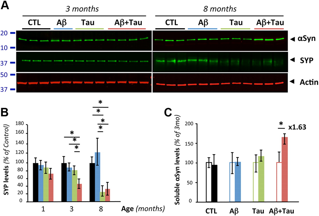
Synergism between Aβ/APP and tau is required to elevate αSyn protein levels in vivo.
A, Quantitative Western blot analysis of αSyn, SYP, and actin using soluble brain extracts from 3- and 8-month-old nontransgenic (CTL), Tg2576 (Aβ), rTg4510 (Tau), and Tg2576xrTg4510 (AβxTau) mice. B, Quantification of SYP mirrors neuronal toxicity previously reported in rTg4510. Values represent mean ± SD (n = 3–4 per age per genotype; ANOVA followed by Fisher’s PLSD test). C, Quantification of soluble αSyn protein levels revealed no apparent changes with aging in wild-type, Tg2576, and rTg4510 mice. In contrast, an ∼1.6-fold increase of αSyn was observed in 8-month-old Tg2576xrTg4510 compared with 3-month-old animals. Open and filled bars represent data at 3 and 8 months, respectively. Values represent mean ± SD (n = 3–4 per age per genotype; ANOVA followed by Fisher’s PLSD test).
Discussion
Until recently, the principal focus in the field of neurodegenerative diseases has been to understand the pathogenic contributions of misfolded proteins once aggregates have formed. With the emergence of evidence indicating that soluble forms of Aβ may be highly bioactive in AD (Walsh et al., 2002; Lesné et al., 2006; Shankar et al., 2008), there has been a paradigm shift toward studying soluble disease-associated proteins. Aβ and Tau are both examples of this change, as the deleterious effects of soluble Aβ oligomers as opposed to amyloid plaques (Walsh et al., 2002; Shankar et al., 2008) and of soluble tau molecules as opposed to neurofibrillary tangles (Ramsden et al., 2005; Santacruz et al., 2005) were revealed in the last decade. Here we hypothesized that this principle was not restricted to Aβ and tau but may also apply to αSyn.
Accumulation of soluble αSyn in AD in absence of αSyn pathology
Whereas it is well accepted that αSyn can form inclusions in the form of LB and accumulate as Lewy neurites in brains of subjects with AD, little is known about the potential contribution of soluble, nonfibrillar αSyn to AD-related cognitive decline and AD pathophysiology. The results presented here show that soluble levels of intracellular αSyn, as measured by SDS-PAGE, are elevated by approximately twofold in AD compared with age-matched control ITG, even though these AD subjects had no signs of LB/LN pathology.
In agreement with the twofold increase in intracellular αSyn protein levels, transcriptional expression of the SNCA gene was upregulated by ∼1.7-fold. In contrast to our work, both αSyn protein and mRNA levels were not found altered in a various brain regions of a small subset of AD cases (Quinn et al., 2012). Using randomly selected ROS specimens to address this issue (n = 5 per status per brain region; data not shown), we assessed SNCA mRNA levels by rt-qPCR in the inferior temporal gyrus (region used for this manuscript), the midfrontal gyrus, the inferior parietal gyrus, and subregions the entorhinal cortex and calcarine cortex. We found no differences between the control and AD groups in the entorhinal and the calcarine cortices. Gene expression of SNCAwas downregulated in the midfrontal gyrus (1.57-fold; p = 0.025) and upregulated in the inferior temporal gyrus (2.36-fold; p = 0.034), consistent with the ∼1.7-fold increase in αSyn mRNA in our larger ITG samples. Transcript levels showed a trend toward an increase in the inferior parietal gyrus (2.27-fold; p = 0.062). This elevation in SNCA gene expression is surprising considering that one could have expected αSyn to follow an expression pattern similar to other presynaptic proteins such as synaptophysin, i.e., a decrease compared with controls (Lue et al., 1999). However, it parallels quite well the observations made for Aβ or tau, which demonstrated increases in the expression of soluble forms of these proteins in the absence of fibrillar, deposited amyloid plaques (Lesné et al., 2006) or neurofibrillary tangles (Santacruz et al., 2005), respectively.
Soluble αSyn as a new predictor of AD-associated cognitive impairment
Although identifying an accumulation of soluble αSyn in LB-free brains of subjects with AD was unexpected, we believe its relationship to cognitive dysfunction and the strength of that association are what sets αSyn apart as a potentially novel predictor for AD-associated cognitive decline. As reported previously, soluble Aβ, total soluble tau, and several disease-relevant tau species correlated well with cognitive impairment on some measures of memory. However, compared with Aβ and tau, soluble αSyn displayed the strongest correlation indexes in univariate regression analyses. To evaluate the potential predictive power of soluble αSyn for AD-associated cognitive decline in our well characterized cohort, we created multivariate regression models that revealed that αSyn was the most significant predictor variable among those tested for episodic memory, perceptual speed, and global cognition and the only significant predictor variable for semantic memory and working memory. In mice, transgenic animals in which soluble human αSynWT is overexpressed approximately threefold over normal displayed spatial reference memory deficits. Of note, the degree of cognitive impairment observed was similar to those registered for the transgenic mouse model of AD, J20, at the same age. Collectively, our results suggest that soluble αSyn levels may be a crucial correlate and even modulator of cognition in AD. Replication of these findings in different cross-sectional cohorts as well as initiating longitudinal studies will be required to determine whether changes in soluble αSyn in brain and CSF may precede cognitive impairment in humans.
αSyn-induced synaptotoxicity in end-stage AD
αSyn has been implicated in the pathogenesis of Parkinson’s disease, the Lewy body variant of AD, Lewy body dementia, and some forms of prion diseases. These disorders are referred to as synucleinopathies (Hardy and Gwinn-Hardy, 1998). In AD, many groups have reported the presence of insoluble αSyn aggregates (LB/LN) in close association with amyloid plaques both in animal models and postmortem human brain tissue (Masliah et al., 1996, 2001; Lippa et al., 1998; Clinton et al., 2010). However, the importance and relevance of soluble αSyn to AD-associated memory impairment and AD pathophysiology was essentially unknown before the current work.
We describe here that a twofold elevation of monomeric intracellular αSyn, as measured by SDS-PAGE, is accompanied by a selective decrease in expression of presynaptic vesicle proteins, in a similar fashion as that observed in the asymptomatic transgenic I2.2 line (M. K. Lee et al., 2002) overexpressing human αSynWT (threefold overexpression vs endogenous murine αSyn levels) (Nemani et al., 2010). Moreover, we observed a dissociation of αSyn and synapsin protein composition in synaptic vesicles of subjects with clinical AD who had a twofold elevation of soluble αSyn in the IC fraction. This phenomenon was also detected in DLB brains (Scott et al., 2010) and in primary neurons derived from LB-forming PDGF-h-α-syn:GFP transgenic mice (Scott et al., 2010). However, the observed dissociation in the colocalization between αSyn and synapsin reported here occurred in the absence of detectable αSyn inclusions in either cell bodies or neurites (Fig. 6 and data not shown). Accordingly, we did not detect pS129-αSyn immunoreactivity by confocal imaging or Western blotting (data not shown), which form has been classically associated with fibrillar, aggregated αSyn inclusions (Fujiwara et al., 2002; Chen and Feany, 2005; Waxman et al., 2008). These data contrast with the observation that phosphorylation at S129 was detected in synaptic boutons of hippocampal neurons from PDGF-h-α-syn:GFP mice, where the presence of endogenous synaptic vesicle proteins was altered (Scott et al., 2010). Although the molecular mechanism underlying the accumulation of soluble αSyn in AD neurons remains unknown, it is likely that both the selective decrease in specific presynaptic proteins and the dissociation in the normal protein composition of synaptic vesicles are responsible for an impaired neurotransmitter release by neuronal cells (Nemani et al., 2010; Scott et al., 2010). Our results provide medically relevant support to the notion that an imbalance of endogenous soluble αSyn expression at presynaptic terminals in AD alters its physiological function in regulating neurotransmitter vesicular release (Garcia-Reitböcket al., 2010; Nemani et al., 2010; Scott et al., 2010). Importantly, our findings therefore suggest that dysregulation of soluble αSyn represents a crucial and previously unreported neurobiological event occurring during AD physiopathology.
Therapeutic implications
Although APP transgenic mice are viewed as better models for familial than sporadic AD, Tg2576 (Hsiao et al., 1996) and J20 (Mucke et al., 2000) APP transgenic lines are commonly used for basic and therapeutic (drug development) research. It was therefore relevant to ask whether such mice underwent an abnormal accumulation of soluble αSyn (data not shown for J20). In either line, we did not detect any change in endogenous soluble mouse αSyn across genotypes at the ages of 1, 3, 8, 13, and 17 months. Despite the presence of amyloid plaques in 13- and 17-month-old animals, these findings differ greatly from the described accumulation of soluble αSyn seen in brain tissue from nondominant AD cases, suggesting several possibilities. First, murine (m-αSyn) and human (h-αSyn) αSyn sequences are different, especially in the C-terminal end of the protein. Despite the presence of a threonine at position 53, such as the A53T human mutant linked to PD, and the capability of m-αSyn to undergo profound neuropathological changes when overexpressed in vivo (Rieker et al., 2011), soluble m-αSyn did not accumulate in the APP transgenic mice tested. This suggested that an important cofactor related to αSyn was missing in APP transgenic mice but was present in humans. Of course, tau came to mind. It is well accepted that human tau and αSyn amyloidogenic inclusions can occur in the same cells in human brains (Lee et al., 2004) and that both proteins can potentiate the aggregation of each other in vitro (Giasson et al., 2003b) and in vivo (Clinton et al., 2010). It was revealing that in our cohort, intercorrelation analyses between the respective levels of soluble forms of tau and αSyn not only support the concept just mentioned but also advance the notion that the pro-amyloidogenic properties of these proteins can be extended to their respective soluble monomeric forms in vivo (Table 3). This seemed to be specific to measures of total tau and pS202-Tau but not to Alz50-Tau (more associated with insoluble PHF), suggesting that the “pas de deux” orchestrated by soluble αSyn and soluble tau might represent an early event in AD pathogenesis. Because we did not observe the presence of fibrillar or pS129-αSyn-reactive lesions in the AD brains with elevated soluble αSyn, we believe our data relate to the second predicted model of interaction presented by Lee et al. (2004). In this model, native αSyn can interact with preamyloid tau species (possibly pS202-Tau as indicated by our intercorrelation analyses), facilitating the conversion of αSyn into a preamyloidogenic molecule. Our mouse genetic data support this concept but also indicate that overexpression of human Aβ/APP or tau alone was not sufficient to drive the increase in αSyn expression. Instead our results suggest that a synergy between Aβ/APP and tau is required to abnormally elevate soluble αSyn levels. Whether or not this preamyloidogenic form of αSyn corresponds to a specific oligomeric assembly of αSyn present in neurons (Bartels et al., 2011) remains to be determined.
In conclusion, our data suggest that AD may not be a disease of two misfolded proteins but instead might result from at least a three-pronged attack on neurons by soluble Aβ, tau and αSyn. We propose a model in which soluble Aβ species alters soluble tau function and chemistry, thereby allowing soluble αSyn to accumulate. This deleterious elevation of αSyn then leads to a selective decrease in certain presynaptic vesicle proteins and a dissociation of the protein composition of these vesicles, impairing neurotransmitter release at synapses already under siege by soluble Aβ oligomers and abnormal tau species at postsynaptic sites. Thus, the studies reported here document a crucial and previously undocumented feature of AD pathophysiology.