
Progesterone Receptor Membrane Component-1 May Promote Survival of Human Brain Microvascular Endothelial Cells in Alzheimer’s Disease

By Xin Xu, Xiangyan Ruan, Rui Ju, Zhikun Wang, Yu Yang, Jiaojiao Cheng, Muqing Gu, and Alfred O. Mueck
Excerpt from the article published in American Journal of Alzheimer’s Disease & Other Dementias. 2022. Published June 22, 2022 DOI:10.1177/15333175221109749
Editor’s Highlights
- Cerebral amyloid angiopathy (CAA) is found in the majority of Alzheimer’s disease (AD) cases.
- The vascular accumulation of β-amyloid (Aβ) is the causative agent of CAA, with the characteristics of dysfunctional hemodynamics and leaky blood-brain barrier (BBB).
- Progesterone receptor membrane component 1 (PGRMC1) is a protein of the MAPR family which binds Aβ oligomers in AD brain tissue and this interaction mediates synaptotoxic effects of Aβ.
- PGRMC1 overexpression enhanced the proliferation of human brain microvascular endothelial cells (HBMVECs), migration, and angiogenesis.
- PGRMC1 was found to be low expressed in the frontal cortex and temporal cortex tissues of Alzheimer’s disease.
Abstract
Cerebrovascular changes occur in Alzheimer’s disease (AD). The progesterone receptor membrane component-1 (PGRMC1) is a well identified hormone receptor with multiple functions in AD. This study aims to explore the involvement of PGRMC1 in the regulation of vascular endothelial function, providing new therapy options for AD. Single-cell sequencing revealed that the expression of PGRMC1 is lower in AD. By bioinformatics analysis, we found PGRMC1 was associated with regulation of cell proliferation, angiogenesis and etc. To understand the functional significance of PGRMC1, knockdown and overexpression were performed using human brain microvascular endothelial cells (HBMVECs), respectively. Cell proliferation assay, migration assay, tube formation assay were performed in experiments. We demonstrated that the overexpression of PGRMC1 promoted the cellular processes associated with endothelia cell proliferation, migration, and angiogenesis, significantly. In conclusion, PGRMC1 may contribute to the modulation of HBMVECs function in AD. This finding may offer novel targets for AD treatment.
Introduction
Alzheimer’s disease (AD) is the most prevalent cause of dementia, with numbers projected to attain 115.4 million worldwide by 2050.1,2 β-amyloid (Aβ) and hyperphosphorylated tau pathology are major pathological hallmarks of AD.3 Furthermore, cerebrovascular dysfunction plays a critical role in the earliest pathogenesis of AD. As a strong contributor to vascular dysfunction, Cerebral amyloid angiopathy (CAA) is found in the majority of AD cases. The vascular accumulation of Aβ is the causative agent of CAA, with the characteristics of dysfunctional hemodynamics and leaky blood-brain barrier (BBB). CAA ultimately leads to clearance failure and further accumulation of Aβ in the cerebrovasculature and brain parenchyma.4,5 The perivascular deposition of Aβ in the brain leads to endothelial cell dysfunction and death.6
Progesterone receptor membrane component 1 (PGRMC1) is a protein of the MAPR family which is involved in multiple functions, including cytochrome P450 regulation,7 autophagy, membrane trafficking,8 fertility,9 steroidogenesis,10 and AD.11,12 In central nervous system, progesterone has been demonstrated to exert neurogenic and neuroprotective effects via PGRMC1. It has further been shown that PGRMC1 binds Aβ oligomers in AD brain tissue and that this interaction mediates synaptotoxic effects of Aβ.13 However, the role of PGRMC1 in endothelia cell especially in AD has not been investigated yet. It has been demonstrated PGRMC1 promotes luteal vascularization in mice.14 PGRMC1 might also contribute to VEGF-induced neovascularization in tumor tissue.15
In the present study, we investigated the expression of PGRMC1 in AD and explored the role of PGRMC1 in human brain microvascular endothelial cells (HBMVECs) by bioinformatic analysis and experimental verification.
Results
Progesterone Receptor Membrane Component-1 is Downregulated in Alzheimer’s Disease
To investigate the potential role of PGRMC1 in AD, we first analyzed the expression level of PGRMC1 in AD and NC, and found that the transcripts of PGRMC1 in AD samples was dramatically decreased compared with the NC samples from both frontal cortex and temporal cortex tissues (Figure 1A).
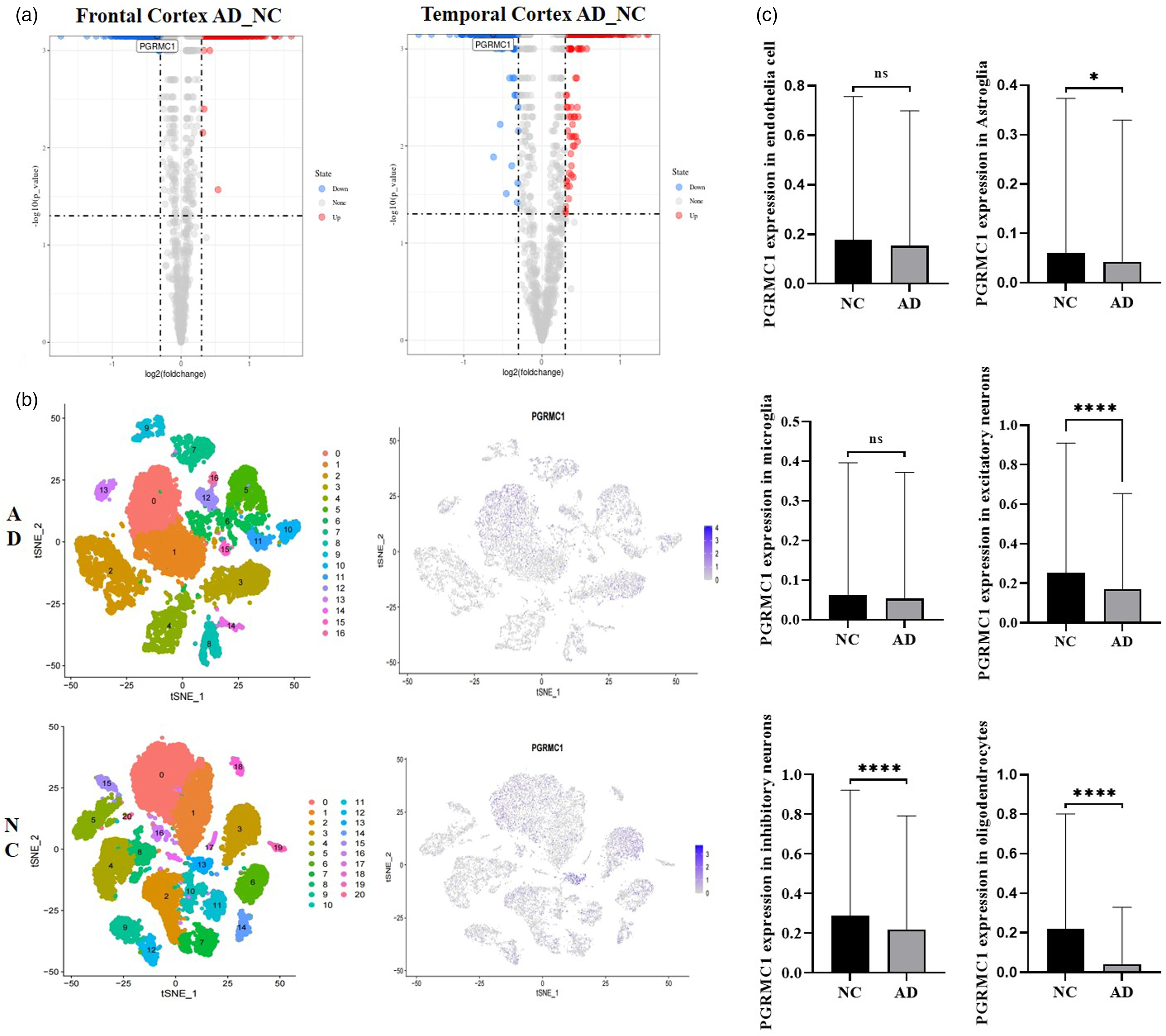
(A) Volcano plots showing DEGs between alzheimer’s disease and normal control in frontal cortex and temporal cortex, respectively. (B) Single-cell RNA sequencing analysis and the expression of PGRMC1 in single-cell dataset GSE157827. (C) Expression of PGRMC1 in different kinds of cells from single-cell dataset GSE157827 ns P > .05 *P < .05, ****P < .0001. Abbreviations: PGRMC1, progesterone receptor membrane component-1.
To establish a baseline profile of cell populations, t-SNE visualization of cell types isolated from the AD and NC samples, with yielded 36 unique cell clusters (AD: 16; NC: 20). Once cell clusters have been identified, these are interpreted according to marker genes from Shun-Fat Lau’s study16 (Figure 1B). There are six kinds of cells: astrocytes (AQP4, ADGRV1, GPC5, and RYR3), endothelial cells (CLDN5, ABCB1 and EBF1), excitatory neurons (CAMK2A, CBLN2 and LDB2), inhibitory neurons (GAD1, LHFPL3 and PCDH15), microglia (C3, LRMDA and DOCK8), and oligodendrocytes (MBP, PLP1 and ST18). Violin plots showed the expression of representative marker genes across different clusters in both groups (Supplementary Figures 1–12). PGRMC1 exhibited a decrease in AD endothelial cells, microglia, astrocytes, excitatory neurons, inhibitory neurons, and oligodendrocytes (Figure 1C). In the latter four kinds of cells, comparisons were statistically significant. To assess the function of PGRMC1 in AD endothelial cells, we divided ECs in AD to two groups according to PGRMC1 expression. (High PGRMC1-expression group: > 0; Low PGRMC1-expression group: = 0) The differentially expressed gene (DEG) PPI network was comprised 1291 nodes and 28036 edges, and the subnetwork was acquired through selecting the DC which was higher than 60, including 289 nodes and 11315 edges (Figure 2A). Based on the subnetwork, the enriched gene function was highly correlated with regulation of cell proliferation, angiogenesis and etc. (Figure 2B).
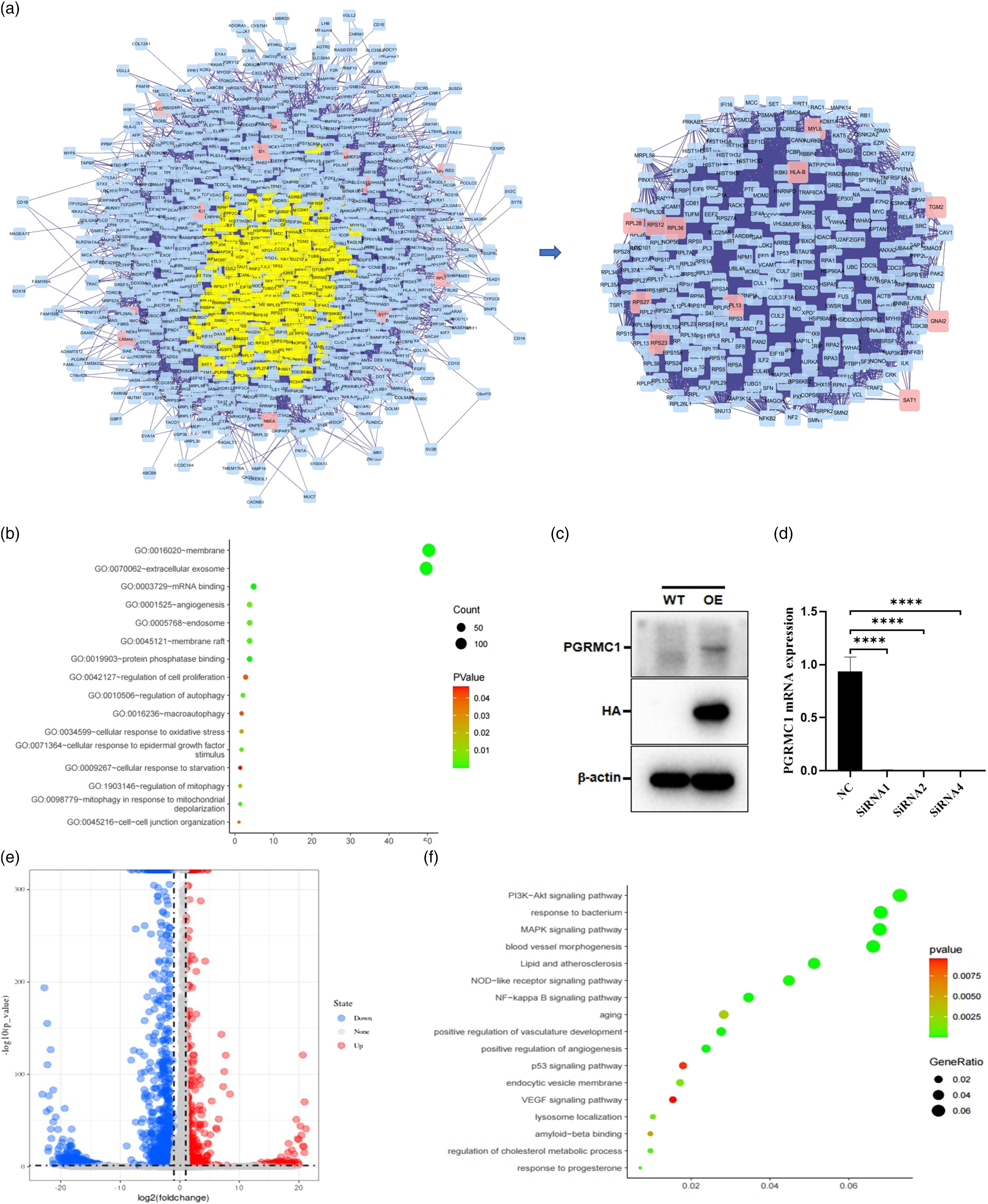
(A) PPI construction network and topological analysis of DEGs in between low- and high-PGRMC1 expression endothelial cells. (B) Functional enrichment of DEGs between low- and high-PGRMC1 expression endothelial cells. (C) Western blot results showing PGRMC1 was overexpressed in HBMVECs. (D) Real-time PCR results showing the efficiency of PGRMC1 siRNA transfection in HBMVECs. **** P < .0001. (E) Volcano plot showing the fold change and significance of gene expression values between PGRMC1-overexpressing and control HBMVECs. (F) Functional enrichment of DEGs between PGRMC1-overexpressing and control HBMVECs. Abbreviations: PGRMC1, progesterone receptor membrane component-1; HBMVECs, human brain microvascular endothelial cells.
To understand the roles of PGRMC1 in the development of AD, we attempted to overexpress and knockdown PGRMC1 in HBMVECs. First, HBMVECs which stably expresses PGRMC1 was constructed by lentivirus infection and puromycin selection. The protein expression of PGRMC1 was verified by Western blot. As shown in Figure 2C, PGRMC1 was overexpressed compared with control vector. siRNA sequences knocked down PGRMC1 mRNA levels as determined through qPCR in Figure 2D. To validate the above results, we performed the high-throughput sequencing for above PGRMC1 stable cell lines and control. Differential gene expression analysis (P < .05, 2-fold threshold) revealed significant change in 578 (up) and 1559 (down) genes (Figure 2E). Functional enrichment analysis revealed that DEGs were highly correlated with PI3K-Akt signaling pathway, Lipid and atherosclerosis, aging, positive regulation of angiogenesis, VEGF signaling pathway, lysosome localization, amyloid-beta binding, regulation of cholesterol metabolic process, response to progesterone (Figure 2F).
Progesterone Receptor Membrane Component-1 Promotes Human Brain Microvascular Endothelial Cells Proliferation, Migration and Tube Formation
Endothelial proliferation, migration, and tube formation in response to VEGF stimulations are key processes during angiogenesis. To explore the effects of PGRMC1 on HBMVECs, we generated HBMVECs stably expressing PGRMC1. In this study, we demonstrated that PGRMC1 overexpression prominently promoted migration, 48 hours later (P < .05) (Figure 3A, B and E). Correspondingly, PGRMC1 knockdown inhibited cell migration (P < .01) (Figure 3C, D and F). We also found that the overexpression of PGRMC1 significantly promoted proliferation in vitro (Figure 3G). Moreover, overexpressing PGRMC1 in HBMVECs resulted in a significant increase in vessel structures (Figure 4A and B). While, PGRMC1 knockdown inhibited VEGF-induced angiogenesis 12 hours later (Figure 4C and D). The total tube length in the control cells was significantly shorter than those in the PGRMC1 overexpressing cells (P < .05). And the total tube length in the PGRMC1 knockdown cells was significantly shorter than those in NC cells (Figure 4E) (P < .05). Similarly, PGRMC1 overexpressing cells have higher number of branch points than the control group (P < .05) and PGRMC1 knockdown cells (P < .0001), significantly (Figure 4F). Altogether, the above results demonstrated PGRMC1 promotes HBMVECs proliferation, migration and tube formation.
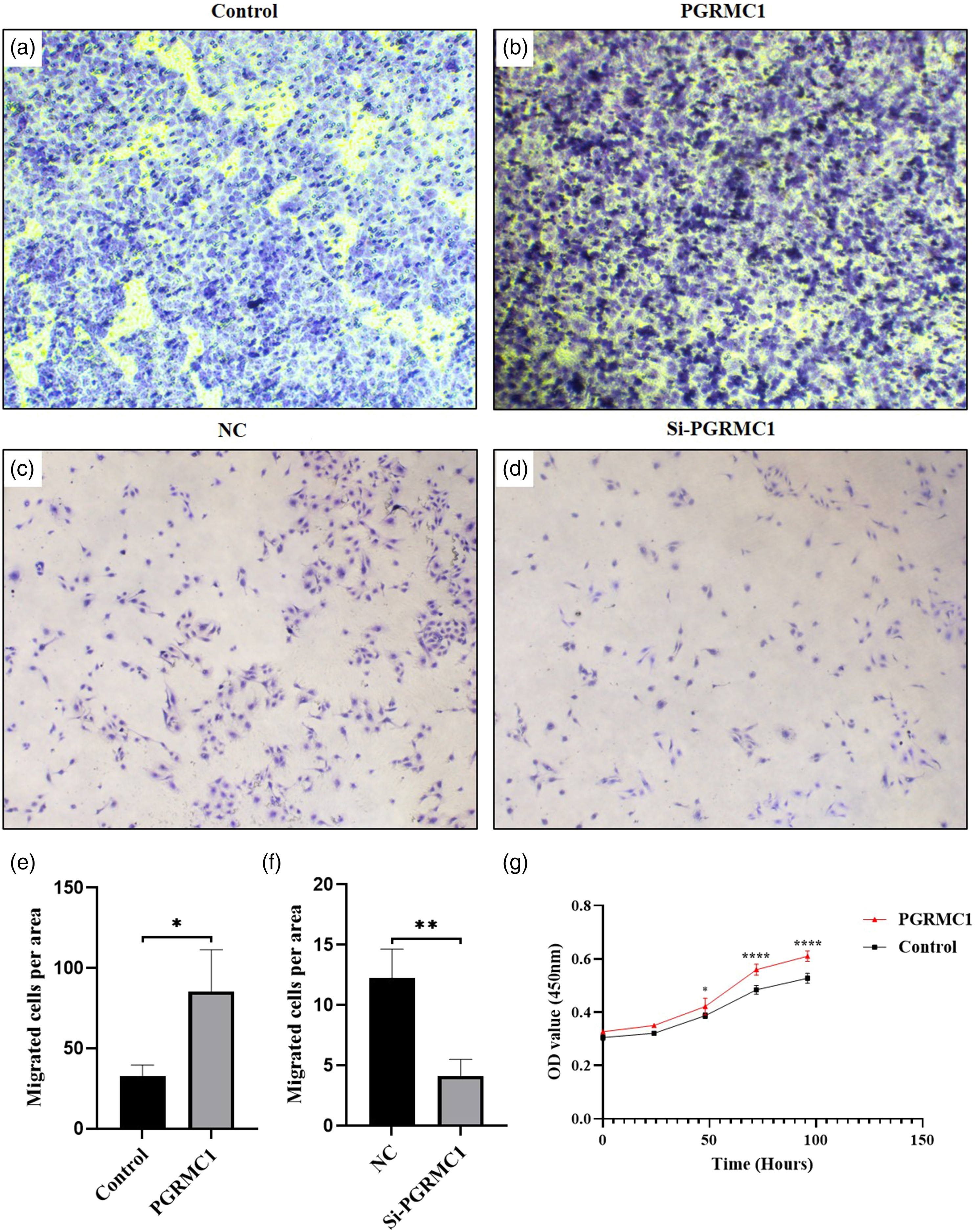
Progesterone receptor membrane component-1 promotes cell migration and cell proliferation in vitro. (A-D) Representative images of cells that migrated through the membranes after 48 hours. (magnification ×40). (E-F) The numbers of cells that migrated through the membranes after 48 hours. Data were presented as mean ± SD. The results were reproducible in three independent experiments. (G) The viability of human brain microvascular endothelial cells were tested by CCK-8 assay. *P < .05, **P < .01, ****P < .0001.
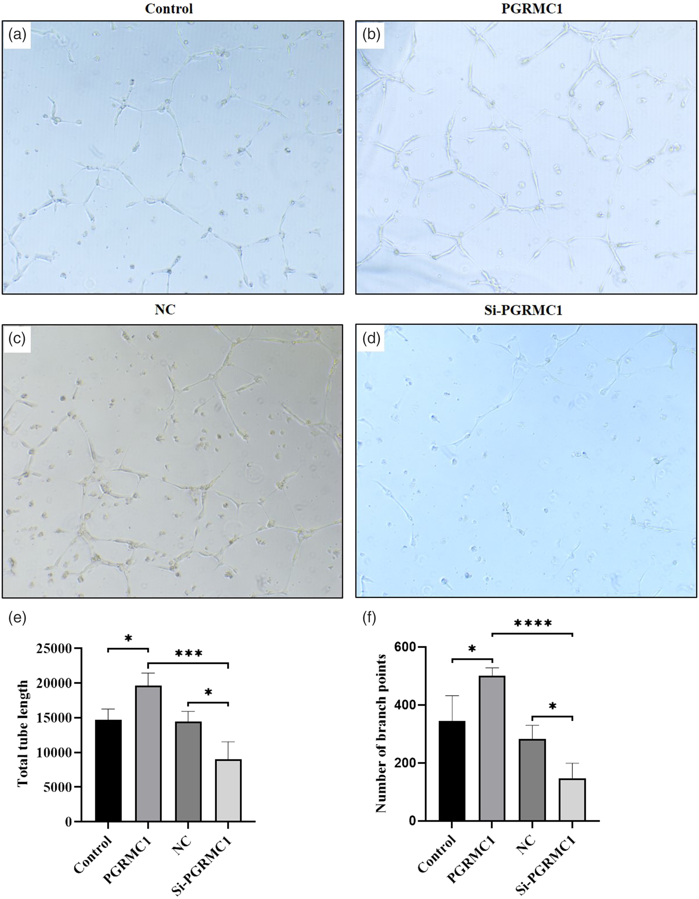
Progesterone receptor membrane component-1 promotes angiogenesis in vitro. (A-D) Representative images of angiogenic response after 12 hours. (magnification ×100). (E) The number of branch points of angiogenic response after 12 hours. (F) Total tube lengths of angiogenic response after 12 hours. Data were presented as mean ± SD. The results were reproducible in four independent experiments. *P < .05, **P < .01, ***P < .001, ****P < .0001.
Discussion
As the most common cause of dementia, AD severely affects the life quality of elderly patients. Currently, no effective medications are available for AD, because of the limited understanding of its pathophysiological mechanism.19 Recent researchs demonstrated that frontal periventricular and posterior white matter hyperintensities are associated with CAA in familial AD.20 Vascular amyloid deposits could be early events in AD and lead to decreased Aβ clearance, and, finally, lead to neurodegeneration.21 However, new biomarkers and therapeutic targets for CAA in AD are urgently needed.
Previous studies have reported that the PGRMC1 played an important role in the nervous system, especially in the pathological mechanism of AD. Its possible molecular pathways involved in AD include binding to Aβ oligomers, regulation of Ca ion homeostasis, glial cell BNDF secretion, nerve cell apoptosis, nerve cell glucose metabolism, Ras signaling pathway, cholesterol metabolism, etc.11,12,22 In the present study, for the first time, the expression level of PGRMC1 in single-cell RNA-sequencing data of AD were explored. Based on the analysis of transcriptome data of Shun-Fat Lau’s study,16 PGRMC1 was found to be low expressed in the frontal cortex and temporal cortex tissues of Alzheimer’s disease. We next detected the expression of PGRMC1 in different cell types. The results showed a downward trend in all kinds of cells in AD, including endothelial cells, microglia, astrocytes, excitatory neurons, inhibitory neurons, and oligodendrocytes. Although the decrease in endothelial cells did not reach statistical significance, we found that the DEGs associated with PGRMC1 were highly enriched in regulation of cell proliferation and angiogenesis. Similar findings have been demonstrated in our HBMVECs overexpressed PGRMC1. PGRMC1 overexpression enhanced the proliferation of HBMVECs. Prior studies that have noted the importance of PGRMC1 in cell proliferation and metastasis, such as breast cancer, oral cancer.23,24 What is surprising is that, we demonstrated that PGRMC1 promoted HBMVECs tube formation. To date, few studies have reported the role of PGRMC1 in angiogenesis. Nevertheless, the detailed mechanism remains to be determined yet.
Conclusion
Our findings identified PGRMC1 as a critical gene for AD and revealed its involvement in proliferation, migration, and angiogenesis of HBMVECs. Further study on pathogenesis and hormone might provide new insight into the potential treatment of AD.