
Nuclear and cytoplasmic huntingtin inclusions exhibit distinct biochemical composition, interactome and ultrastructural properties

By Nathan Riguet, Anne-Laure Mahul-Mellier, Niran Maharjan, Johannes Burtscher, Marie Croisier, Graham Knott, Janna Hastings, Alice Patin, Veronika Reiterer, Hesso Farhan, Sergey Nasarov, and Hial A. Lashuel
Excerpt from the article published in Nature Communications, volume 12, Article number: 6579 (2021) DOI: https://doi.org/10.1038/s41467-021-26684-z
Editor’s Highlights
- Huntington’s disease (HD) is an autosomal dominant genetic and progressive neurodegenerative disorder caused by the abnormal expansion of CAG trinucleotide repeats within exon 1 of the huntingtin gene (HTT).
- The expanded repeats are translated into a long pathogenic polyglutamine (polyQ) tract (> 36 repeats) that renders the Huntingtin protein (Htt) more prone to aggregate.
- The length of the CAG repeat is inversely correlated to the age of onset, with the juvenile form associated with a polyQ repeat length of 75 or more.
- Incomplete splicing of HTT leading to the overexpression of N-terminal fragments of mutant Htt comprising the Exon 1 region (Httex1), which contains the polyQ expansion has been shown to occur in HD patient’s brains, and Httex1 protein was previously described as a key component of the intracellular inclusions found in HD post-mortem brains.
- Expression of Httex1 72Q in primary cortical neurons leads to the formation of dense and filamentous nuclear inclusions.
- Only tag-free Httex1 72Q overexpression results in mitochondrial fragmentation and is characterized by the strong accumulation of inflammation-linked proteins in the aggregates.
- modifiers of Htt inclusion growth and aberrant secondary interactions with other proteins and organelles represent an alternative and strategy for interfering with Htt-induced toxicity and slowing disease progression, especially after disease onset.
Abstract
Despite the strong evidence linking the aggregation of the Huntingtin protein (Htt) to the pathogenesis of Huntington’s disease (HD), the mechanisms underlying Htt aggregation and neurodegeneration remain poorly understood. Herein, we investigated the ultrastructural properties and protein composition of Htt cytoplasmic and nuclear inclusions in mammalian cells and primary neurons overexpressing mutant exon1 of the Htt protein. Our findings provide unique insight into the ultrastructural properties of cytoplasmic and nuclear Htt inclusions and their mechanisms of formation. We show that Htt inclusion formation and maturation are complex processes that, although initially driven by polyQ-dependent Htt aggregation, also involve the polyQ and PRD domain-dependent sequestration of lipids and cytoplasmic and cytoskeletal proteins related to HD dysregulated pathways; the recruitment and accumulation of remodeled or dysfunctional membranous organelles, and the impairment of the protein quality control and degradation machinery. We also show that nuclear and cytoplasmic Htt inclusions exhibit distinct biochemical compositions and ultrastructural properties, suggesting different mechanisms of aggregation and toxicity.
Introduction
Huntington’s disease (HD) is an autosomal dominant genetic and progressive neurodegenerative disorder caused by the abnormal expansion of CAG trinucleotide repeats within exon 1 of the huntingtin gene (HTT)1,2,3,4. The expanded repeats are translated into a long pathogenic polyglutamine (polyQ) tract (> 36 repeats) that renders the Huntingtin protein (Htt) more prone to aggregate5. The length of the CAG repeat is inversely correlated to the age of onset, with the juvenile form associated with a polyQ repeat length of 75 or more6,7. The accumulation of Htt-positive intraneuronal aggregates and inclusions in the cortex and the striatum of post-mortem brain from HD patients and in animal and cellular models of HD2 has led to the hypothesis that Htt aggregation plays a central role in the pathogenesis of HD. However, our understanding of the ultrastructural and biochemical composition of Htt inclusions and the molecular mechanisms that drive their formation, clearance, and toxicity remains incomplete. Addressing this knowledge gap is crucial to understand the molecular and cellular mechanisms underpinning HD and to enable the development of effective therapies and disease-modifying strategies for this disease.
Although several studies have investigated specific cellular mechanisms linked to Htt aggregation, the current models often lack detailed characterization of the inclusions at both the biochemical and structural levels. Furthermore, while some of the previous studies explored the effect of polyQ repeat length on the ultrastructure properties of mutant Htt, there are no reports on the role of the first N-terminal 17 amino acids (Nt17) of Htt – which regulates many aspects of the aggregation and cellular properties of Htt proteins8,9,10,11 – on the organization and ultrastructure properties of cytoplasmic and nuclear Htt inclusions. Finally, the vast majority of the previous studies used mutant Htt constructs fused to either peptide-based tags or large fluorescent proteins like GFP that could interfere with the cellular and aggregation properties of Htt12,13,14,15.
Here, we employed cellular and neuronal models of Htt cytoplasmic and nuclear inclusion formation to gain insight into how sequence modifications influence the final ultrastructural and biochemical properties of cytoplasmic and nuclear Htt inclusions and their impact on cellular organelles and functions. The cellular models used in this study are based on the overexpression of N-terminal fragments of mutant Htt comprising the Exon 1 region (Httex1), which contains the polyQ expansion. Incomplete splicing of HTT leading to Httex1 protein expression has been shown to occur in HD patient’s brains16, and Httex1 protein was previously described as a key component of the intracellular inclusions found in HD post-mortem brains16,17,18. Moreover, the expression of pathogenic Httex1 (polyQ tract > 43Q) is sufficient to induce HD-like features, including aggregates formation and toxicity in mice19,20,21, Drosophila22, C. elegans23 and cell culture models11,12,15,24,25,26,27,28,29,30,31. In vitro and cellular studies also showed that Httex1 aggregates in a polyQ repeat length and concentration-dependent manner32,33,34. Therefore the mutant Httex1-based models are useful to study the pathogenesis of HD as they reproduce different aspects of Htt aggregation and have been instrumental in advancing our understanding of the sequence, molecular, and structural determinants of Htt aggregation and inclusion formation10,26,27,28,29,35,36.
We applied a combination of Correlative Light and Electron Microscopy (CLEM) and proteomics-based approaches to investigate the structural and biochemical properties of the cytoplasmic and nuclear Httex1 inclusions in HEK 293 cells and primary cortical neurons. We also investigated the role of the Nt17 domain and the polyQ tract length in modulating the composition and the structural and toxic properties of mutant Httex1 inclusions. Finally, given that a large body of published cellular studies on the mechanisms of Httex1 aggregation and inclusion formation is based on constructs in which Httex1 is fused to the GFP9,11,12,37,38,39,40, we also compared, for the first time, the composition, ultrastructural properties, and toxicity of inclusions formed by native (tag-free) and GFP-tagged mutant Httex1 proteins.
Our results demonstrate that Htt inclusions are composed of a complex mixture of aggregated mutant Httex1, different cellular proteins and membranous organelles, including the endosomal system. We show that functional and ultrastructural properties of Httex1 inclusions are differentially altered by sequence modifications and interactions with lipids and cellular organelles. We demonstrate that Htt cytoplasmic and nuclear inclusions exhibit distinct composition and ultrastructural properties, suggesting different mechanisms of aggregation, inclusion formation, and toxicity. These observations revealed that the differences in the cellular environment and interactome could influence the mechanisms of aggregation, the structural and biochemical properties of the inclusions, and their relative contribution to mutant Htt-induced toxicity. Finally, we show that inclusions produced by mutant Httex1 72Q-GFP exhibit striking differences in terms of organization, ultrastructural properties, protein composition, and their impact on mitochondrial functions compared to the inclusions formed by the tag-free mutant Httex1 72Q. Overall, our findings contribute to a better understanding of the sequence, molecular, and cellular determinants of mutant Httex1 aggregation, inclusion formation, and toxicity. They also underscore the critical importance of further studies to investigate the role that lipids, organelles, and the Htt interactome play in Htt pathology formation and maturation, and Htt-induced toxicity in HD.
Results
Httex1 cytoplasmic inclusions exhibit a distinctive core and shell morphology and are composed of highly organized fibrils, cytoplasmic proteins, and membranous structures
To investigate the ultrastructural properties of Htt inclusions, we first used a mammalian cell model system of HD, in which overexpression of mutant Httex1 with polyQ repeats > 39 has been shown to result in a robust and reproducible formation of cytoplasmic Htt inclusions15,29. In this model, nuclear inclusions are also observed in ~15% of transfected cells by Httex1 72Q41, thus providing an opportunity to investigate and compare the ultrastructural features of cytoplasmic and nuclear Htt inclusions under identical conditions. This model system is widely used to investigate molecular, cellular, and pharmacological modulators of Htt aggregation and inclusion formation11,12,15,24,25,26,27,28,29,30,31,42,43, though very often using Htt constructs with extra non-natural sequences such as Myc, FLAG, and GFP tags. Previous studies have shown that the presence of such tags significantly alters the aggregation properties of N-terminal fragments of mutant Htt44,45,46. Therefore, in this work, we opted to maintain the native sequence of the protein and investigated mutant Httex1 aggregation and inclusion formation in the absence of additional sequences or tags. Tag-free Httex1 72Q was overexpressed in HEK 293 cells (HEK cells), and the morphology and structural properties of Httex1 inclusions were assessed. Despite the formation and abundance of inclusions formed by Httex1 72Q, we did not observe overt toxicity in HEK cells. The initiation of apoptotic events was apparent only after 96 h, as indicated by Caspase 3 activation without loss of plasma membrane integrity41.
Next, we assessed the morphology of Httex1 72Q inclusions by immunocytochemistry (ICC) using several antibodies against different epitopes along the sequence of Httex1 (Fig. 1a and S1). Interestingly, all the antibodies presented a strong immunoreactivity to the periphery of the Httex1 72Q inclusions, and none of these antibodies labeled the core of these inclusions (Fig. 1b and S2a). This observation suggests either an absence of Htt in the center of the inclusions or poor accessibility of the used antibodies to the core of the inclusions, possibly due to the high compactness of the Htt aggregates in the core compared to the periphery of the inclusions. In addition, we observed high colocalization of the filamentous actin (visualized by phalloidin) with Httex1 inclusions (Fig. 1b, white arrowheads), indicating a possible involvement of the cytoskeleton proteins in Htt inclusion formation.
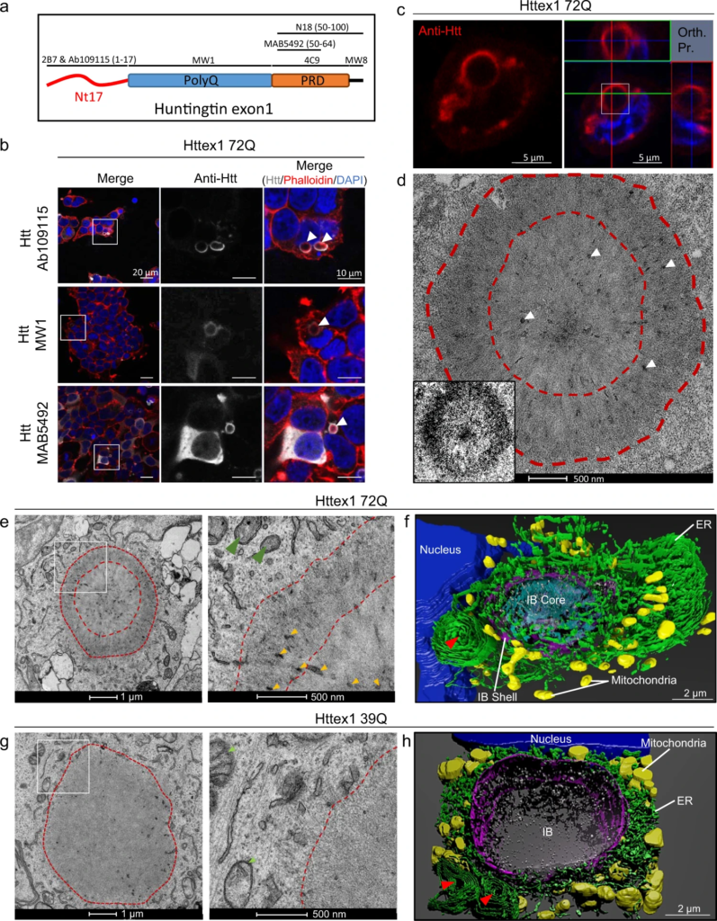
Confocal microscopy and CLEM revealed the ring-like structure of the Httex1 72Q inclusions in HEK cells.
a Epitope mapping of the Httex1 antibodies used in this study. b Confocal imaging of Httex1 72Q inclusions formed 48 h after transfection in HEK cells. All the Htt antibodies showed strong immunoreactivity to the periphery of the inclusions. The nucleus was counterstained with DAPI and the F-actin with phalloidin. White arrows indicate the colocalization of the F-actin with the ring-like structure of Httex1 inclusions. Scale bar = 20 μm and 10 μm. c, d CLEM of Httex1 72Q inclusions. c Confocal imaging of Httex1 72Q inclusions formed 48 h after transfection in HEK cells. The selected area (white square) was examined by EM (d). Orthogonal projection (Orth. Pr.), Scale bars = 5 μm. d The presence of membrane fragments and vesicles are indicated by the white arrowheads. The binary image (inset) shows the core and the shell ultrastructure of the Httex1 72Q inclusion. Scale bar = 500 nm. e Representative EM images of Httex1 72Q inclusion. Higher magnification (white square) are shown in the right panel. Dashed lines delimit the aggregate and the core of the inclusion. Internalized membranous structures and the mitochondria are indicated by the orange and the green arrowheads, respectively. Scale bar = 1 μm and 500 nm. f 3D model of Httex1 72Q cellular inclusion and surrounding organelles (top view). The Httex1 inclusion body (IB) shell is represented in purple, the core in cyan. ER membranes are shown in green, intra-inclusion membranous structures in white, the nucleus in blue, mitochondria in yellow, and the stacked ER cisternae are indicated by a red arrowhead. Scale bar = 2 μm. g Representative EM images of Httex1 39Q inclusion formed 48 h after transfection in HEK cells. The white square indicates the area shown in the right panel at higher magnification. Dashed lines delimit the inclusion. Scale bar = 1 μm and 500 nm. h 3D model of the Httex1 39Q IB is shown in purple, surrounded by mitochondria (yellow), ER structures (green), stacked ER cisternae (red arrowheads), intra-inclusion membranous structures are shown in white and the nucleus in blue. Scale bar = 2 μm.
To gain more insight into the structural and organizational features of Httex1 inclusions formed in cells, we turned to electron microscopy (EM). We first employed a correlative approach to analyze the ultrastructure of the inclusions by EM and their subcellular environment seen with CLEM (Fig. 1c, d). Httex1 72Q positive inclusions were immunostained and imaged by confocal microscopy (Fig. 1c) and then subjected to serial sectioning for analysis by EM (Fig. 1d). The EM micrographs of the Httex1 72Q inclusions revealed a surprisingly complex morphology characterized by a halo-like structure with a dense core and a heavily stained outer shell. The outer layer of the inclusions contained fibrillar structures that appeared to be tightly packed and radiating from the core of the inclusion. EM-dense cytoplasmic structures were detected in both the core and the periphery and could reflect the incorporation or sequestration of disrupted organelles inside the inclusion12,47,48(Fig. 1d, white arrowheads). The halo-morphology of Httex1 inclusions was consistent across many imaged inclusions (Supplementary Fig. 3a).
To further characterize the structural properties and distribution of Httex1 within the inclusion, we used cryo fixation via high-pressure freezing, which preserves the cellular ultrastructure in its native state. EM imaging revealed radiating fibrils at the periphery and more tightly organized and stacked fibrils in the core (Supplementary Fig. 4a). Electron-dense membranous structures were observed in the core and more to the periphery. These results suggest that both the core and shell contain aggregated fibrillar forms of mutant Htt but with a distinct structural organization. The structural organization of Httex1 72Q inclusions combined with the primary localization of Htt antibodies in their periphery suggests that Htt fibrils in the outer layer of these inclusions serve as active sites for the recruitment of soluble Htt, the growth of Htt fibrils, and the interaction of Htt with other proteins and cellular organelles.
To determine whether the formation of cytoplasmic inclusions involves interactions with or recruitment of membranous structures such as ER and mitochondria, we imaged inclusion positive cells by EM under conditions that preserve the internal membranes of cellular organelles, i.e., in the absence of detergents commonly used in the ICC procedure. EM images revealed that the core and periphery of the Httex1 72Q inclusion contained many small membranous structures (Fig. 1e, yellow arrowheads). An ER network and mitochondria were present at the periphery of these inclusions, suggesting that these regions might act as active sites for the recruitment and interaction of soluble Htt with other proteins and cellular compartments during inclusion formation and maturation. Most of the mitochondria surrounding the cytoplasmic Httex1 72Q inclusions exhibited damaged or markedly reduced numbers of cristae (Fig. 1e, green arrowheads). We next generated a 3D model of the inclusions and the organelles in their vicinity (Fig. 1e, f and Supplementary Movie 1). The 3D model confirmed that inclusions are formed in a crowded region with ER and mitochondria all around it. The 3D model also suggested that the electron-dense membranous structures recruited inside the Httex1 inclusions were mostly composed of endomembranes and vesicles (labeled in white), consistent with a previous report12. Interestingly, the ER adopted a specific “rosette-like” or “stacked cisternae” morphology (highlighted by a red arrowhead) in the periphery of some Httex1 72Q inclusions. Finally, it is noteworthy that the mitochondria (labeled in yellow, Fig. 1f) were not detected inside the inclusion but rather at the periphery of the inclusions. Interestingly, despite their proximity to the nucleus, the inclusion did not compromise the nuclear membrane integrity (Supplementary Fig. 3a).
Next, we sought to identify the endomembrane compartments within Httex1 72Q inclusions using a panel of antibodies or dyes labelling intracellular compartments. The mitochondrial (Tom 20 and Mitotracker) and ER (BiP/Grp78) markers were strongly detected near Httex1 72Q cytoplasmic inclusions (Supplementary Fig. 5a–c). The autophagy flux marker, p62, was enriched in the periphery of Httex1 72Q inclusions (Supplementary Fig. 5d). Markers of aggresome formation, such as Vimentin and HDAC6, were also enriched in the periphery and in close proximity to Httex1 72Q inclusions (Supplementary Fig. 5e, f). Moreover, when cytoskeletal proteins such as actin and tubulin were overexpressed (fused with RFP), they were observed mainly at the periphery of the Httex1 72Q inclusions (Supplementary Fig. 5g, h). This observation is consistent with the colocalization of F-actin (stained by phalloidin) with Httex1-72Q inclusions (Fig. 1b, white arrowheads). None of the organelles’ markers were detected inside the core of these inclusions (Supplementary Fig. 5), although the EM images clearly showed the presence of membranous structures in the center (Fig. 1e). This confirms the poor accessibility of antibodies and dyes to stain the core of the Httex1 inclusions likely due to their compactness and highlights the importance of using EM to decipher the ultrastructural properties and composition of the pathological inclusions.
Altogether our data suggest that Httex1 aggregates could form at the surface of membranes as suggested by Suoponki and colleagues49, which could promote early aggregation events as well as perturbation of membranous structures and their recruitment into cytoplasmic Httex1 inclusions.
Cytoplasmic and nuclear Httex1 inclusions in HEK cells exhibit distinct ultrastructural properties
Both cytoplasmic and nuclear inclusions have been observed in HD patients’ brains and transgenic mouse models of HD50,51,52. Therefore, we compared the structural and organizational properties of Httex1 72Q inclusions in the cytoplasm (~85%) and nucleus (~15%) of the transfected HEK cells containing inclusions41. Using immunofluorescence-based confocal microscopy, we did not observe significant differences in the size or overall morphology between the Httex1 72Q inclusions in the nucleus and the perinuclear region (Fig. 2a). However, EM clearly showed that the nuclear inclusions formed by the Httex1 72Q were enriched in fibrillar structures (Fig. 2b) but did not exhibit the classical core and shell organization nor did they contain the membranous structures trapped within the cytoplasmic inclusion (Fig. 1d, e). This suggests that the intracellular environment is a key determinant of the structural and molecular complexity of the inclusions and that nuclear and cytoplasmic inclusion formation occurs via different mechanisms.
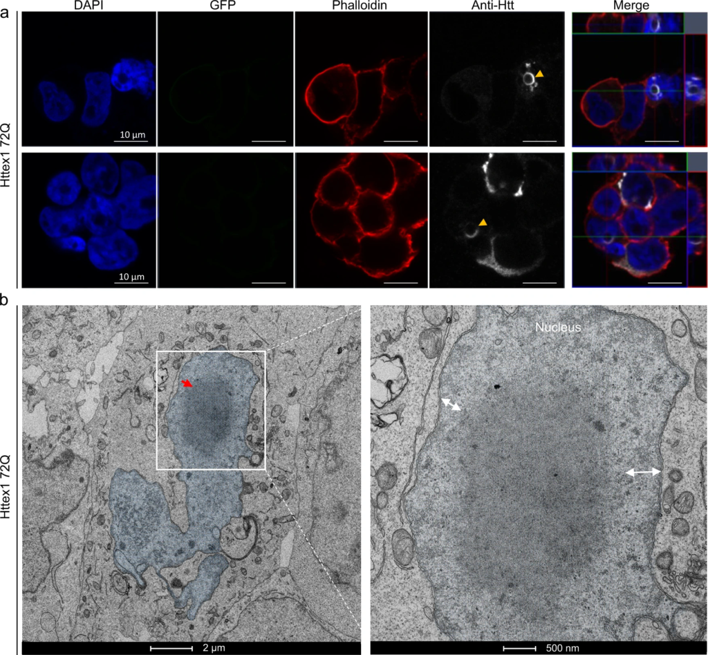
The nuclear inclusions formed by Httex1 72Q do not exhibit the classical core and shell organization observed for the cytosolic inclusions.
a Representative confocal images of Httex1 72Q nuclear inclusions, 48 h after transfection. Httex1 expression (grey) was detected using a specific primary antibody against the N-terminal part of Htt (amino acids 1-17; 2B7 or Ab109115). The nucleus was counterstained with DAPI (blue), and phalloidin (red) was used to stain the actin F. Httex1 nuclear inclusions are indicated by the yellow arrowheads. Scale bars = 10 μm. b Electron micrograph of a representative nuclear Httex1 72Q inclusion. The white square indicates the area shown at higher magnification in the right-hand panel. The nucleus is highlighted in blue, and the double arrows indicate the distance between the nuclear inclusion and the nuclear membrane. No interaction between the nuclear inclusion and the nuclear membrane was observed. Scale bars = 2 μm (left-hand panel) and 500 nm (right-hand panel).
The length of the polyQ domain is another key determinant of inclusion formation
We and others have previously shown that the polyQ repeat length strongly influences the conformation and aggregation properties of Httex1, with higher polyQ favoring the formation of a more compact polyQ domain and accelerating Htt aggregation in vitro and in vivo34,45,53. However, the polyQ dependence on Htt inclusion formation has predominantly been assessed mainly by ICC and in the context of Htt fused to fluorescent proteins and polyQ repeats much longer than the pathogenic threshold (64Q-97Q12; 64Q-150Q30; 43Q-97Q14). Therefore, we next investigated whether the length of the polyQ repeat also influences the organization and ultrastructural properties of cytoplasmic Htt inclusions in HEK cells. Toward this goal, we investigated the level of aggregation and the structural organization of the Httex1 inclusions carrying different polyQ lengths (16Q, 39Q vs. 72Q). Consistent with previous data from our group, no significant cell death was observed in HEK cells overexpressing Httex1 16Q or 39Q constructs even after 96 h41. However, cells expressing Httex1 72Q underwent apoptosis after 96 h. As expected, no inclusions were formed upon overexpression of Httex1 16Q even after 72 h post-transfection (Supplementary Fig. 6). Httex1 39Q inclusions were detected predominantly in the cytoplasm of the HEK cells at all the time points examined (24–72 h), though at lower numbers than in the Httex1 72Q conditions: Httex1 39Q (16%) vs. Httex1 72Q (38%) of transfected cells41.
The dark shell structure that delimits the core from the periphery of the Httex1 72Q inclusions (Fig. 1d, e) was absent in the Httex1 39Q inclusions (Fig. 1g). In addition, the Httex1 39Q inclusions appeared less dense compared to those of the Httex1 72Q. These observations were consistent for all eight inclusions imaged per condition (Supplementary Fig. 3b).
Similar to what we observed for Httex1 72Q, the 3D reconstruction of the Httex1 39Q inclusions clearly showed alteration of the ER organization, as well as the localization of mitochondria near the inclusions (Fig. 1h and Supplementary Movie 2). The electron-dense membranous structures found inside the inclusions were identified as endomembranes and vesicles. Httex1 39Q expressing cells also contained specific ER-cisternae at the periphery of the inclusions (Fig. 1h, red arrowheads). Altogether, our data establish that polyQ expansion plays a critical role in determining the final architecture and ultrastructural properties of the Httex1 inclusions.
Removal of the Nt17 domain reduces mutant Httex1 aggregation but does not influence the organization and ultrastructural properties of the inclusions
The Nt17 domain functions as a Nuclear Export Signal (NES) and has been shown to play an important role in regulating the intracellular localization of Htt as well as its aggregation kinetics and extent of inclusion formation36,54. Therefore, we sought to assess the role of Nt17 in regulating the ultrastructural properties of Htt inclusions. Towards this goal, we generated Httex1 39Q and 72Q mutants lacking the entire Nt17 domain (∆Nt17) and compared the structural properties of the inclusions formed by these mutants to those formed by Httex1 39Q and Httex1 72Q. Quantitative confocal microscopy revealed a strong reduction in the number of inclusions (~50% reduction) of cells transfected by Httex1 ∆Nt17 72Q compared to Httex1 72Q41. Surprisingly, inclusions formed by the Httex1 ∆Nt17 72Q (Supplementary Fig. 7a, b) exhibited an architecture and organization (central core and peripheral shell) similar to those formed by Httex1 72Q (Fig. 1d, e and Supplementary Fig. 8a). Furthermore, similar to Httex1 39Q, the Httex1 ∆Nt17 39Q cytoplasmic inclusions did not exhibit a core and shell architecture. These observations suggest that the Nt17 domain – while playing a crucial role in regulating the kinetics and early events of Htt aggregation – does not influence the morphology or structural organization of Htt cytoplasmic and nuclear inclusions. This is surprising given that in vitro and cellular studies have consistently shown that the Nt17 domain plays an important role in regulating the kinetics and structural properties of Htt aggregation11,41 and Htt interactions with lipids and membranes8,9,55,56,57. In a recent study using solid-state nuclear magnetic resonance spectroscopy (ssNMR), Boatz et al. showed that the Nt17 domain and part of the PRD (PPII helices) are buried in the core of the fibrils, while the other part of the PRD (random coil) remains dynamic, accessible and regulates multifilament assemblies in vitro58. Together, these observations could explain why the deletion of the Nt17 domain does not interfere with the organization of the inclusion and suggest that the interactions between the fibrils and other proteins and/or organelles are most likely mediated by the flexible PRD domain.
Neutral lipids are incorporated into Httex1 cellular inclusions in a polyQ length-dependent manner
Although several studies have shown dysfunction of cholesterol metabolism in various cellular and animal models of HD59,60, the role of lipids in Htt inclusion formation and the lipid composition of cellular huntingtin inclusions remains unknown61,62. To gain further insight into the role of lipids in the formation and structural organization of Httex1 inclusions, we next assessed their presence using fluorescent probes targeting different lipid classes. We did not observe the recruitment of ceramide, cholesteryl ester, or phospholipids into mutant Httex1 inclusions (Supplementary Fig. 9a-c). Interestingly, neutral lipids were not found in the center of Httex1 39Q inclusions (Supplementary Fig. 9d white arrowheads) but were enriched in Httex1 72Q inclusions (Supplementary Fig. 9e). This could contribute to the polyQ length-dependent differences in the ultrastructural properties of the Httex1 inclusions (Fig. 1e, g). Neutral lipids were also detected in nuclear inclusions (Supplementary Fig. 9e, yellow arrowheads). Although the Nt17 domain has been shown to act as a lipid- and membrane-binding domain, neutral lipids were also detected in the Httex1 ΔNt17 72Q inclusions, though not inside Httex1 ΔNt17 39Q inclusions (Supplementary Fig. 10a). These results demonstrate that polyQ-dependent interactions between Htt and neutral lipids play an important role in Httex1 aggregation and the formation of both nuclear and cytoplasmic inclusions. Interestingly, these interactions are not dependent on the Nt17 domain. Whether mutant Httex1 interactions with lipids occur prior to aggregation and help initiate its oligomerization or represent late events associated with the maturation of inclusions remains unknown.
Quantitative proteomics reveals that the formation of Httex1 72Q inclusions involves the active recruitment and sequestration of proteins and organelles
To gain further insight into the biochemical composition of Htt inclusions, we performed quantitative proteomic analysis and compared the differentially abundant proteins between the Urea soluble fractions of HEK cells overexpressing Httex1 72Q or Httex1 16Q (see experimental details, Supplementary Fig. 11a, c). Western Blot analysis of the Urea fraction confirmed the presence of Httex1 72Q within inclusions, while non-pathogenic Httex1 16Q was only detected in the soluble fraction (Supplementary Fig. 11a).
As expected, no proteins were significantly enriched in the insoluble fraction of HEK cells expressing Httex1 16Q compared to those expressing GFP (Supplementary Fig. 11d). In contrast, 377 proteins were significantly enriched in the insoluble fraction of HEK cells overexpressing Httex1 72Q compared to cells overexpressing Httex1 16Q (Fig. 3a). Among these proteins, we identified the endogenous HTT protein (Supplementary Fig. 11e). This suggests that the aggregation process triggered by the overexpression of Httex1 also leads to the recruitment of endogenous HTT protein.
Proteomic analysis of the enriched protein contents found in the insoluble fraction of HEK expressing Httex1 72Q reveals protein aggregation and stress response pathways.
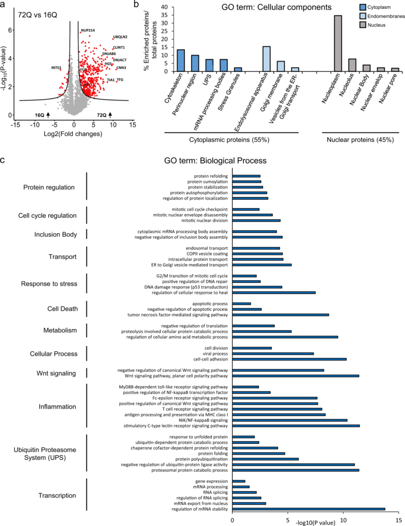
Urea soluble proteins from HEK expressing Httex1 72Q or Httex1 16Q of 3 independent experiments were extracted 48 h after transfection and analyzed using LC-MS/MS. a Identified proteins were plotted using a volcano plot. Black lines represent the threshold of significance at a false discovery rate (FDR) < 0.05 and an S0 of 0.5 which were used for the subsequent analysis. b, c Proteins significantly enriched in Httex1 72Q (red dots in the right part of the volcano plot) were classified by cellular component (b) or biological processes (c) using Gene Ontology (GO) term and DAVID enrichment (−log10(p-value) > 1) analyses. Source data are provided as a Source Data file.
Classification by cellular component (CC, Fig. 3Bb using the Gene Ontology (GO) showed that 55% of the proteins enriched in the insoluble fractions of HEK cells containing Httex1 72Q inclusions were part of the cytoplasmic compartment, with 24% of these proteins belonging to the endomembrane system, including the endolysosomal apparatus (clathrin-coated endocytotic vesicles, early endosome, endosomes, recycling endosomes, exosomes, and autophagosomes), the vesicles involved in Golgi-ER transport, and membranes from the Golgi and trans-Golgi network. These data are in line with the EM data showing that the Httex1 72Q inclusions were composed of small membranous structures and vesicles. Approximately 14% of the proteins enriched in the insoluble fraction were classified as pertaining to the cytoskeleton compartment, with the actin cytoskeleton being the most predominant, consistent with our confocal results (Fig. 1b). The absence of mitochondrial proteins in the soluble Urea fraction confirms that mitochondria are not sequestered inside the inclusions but, rather, accumulate at the periphery, as shown by our EM imaging. The rest of the cytoplasmic proteins sequestered in the insoluble fraction of the Httex1 72Q-transfected HEK cells were mainly from the perinuclear region or from macromolecular protein complexes such as the ubiquitin-proteasome system (UPS), the mRNA processing bodies, and the stress granules. Interestingly, the insoluble fraction of the Httex1 72Q-transfected HEK cells was also significantly enriched by proteins of the nuclear compartment (~45%). Among the nuclear proteins, ~71.6% belonged to the nucleoplasm. Proteins from the nucleolus, the nuclear bodies, the nuclear envelope, and the nuclear pore were also significantly enriched.
Classification of the enriched proteins by biological function showed that the most highly enriched terms for the GO biological process (Fig. 3c) and the signaling pathways using the Ingenuity Pathway Analysis (Supplementary Fig. 12) were related to the proteins involved in the proteasomal ubiquitin-dependent protein degradation. Proteins from the UPS system63, including ubiquitin moiety, E3 ubiquitin ligases (e.g., ITCH, RNF34, TRIM32), 26S proteasome subunits, and deubiquitinases formed the major cluster of the differentially enriched proteins in the Httex1 72Q inclusions. These results are in line with previous studies showing that Htt inclusions formed in HD patients or several cellular and in vivo models sequester several key components of the UPS system, including 26S proteasomes64 (ADRM165), deubiquitinases66 (e.g., NEDD4 and USP5), and E3 ubiquitin ligases (e.g., ITCH, TRAF6, UBE3A, UHRF2, and Parkin) and induce impairment of the UPS67,68,69. In addition, our analysis revealed that among the proteins significantly up- or down-regulated in Httex1 72Q compared to Httex1 16Q, 42 proteins are known Htt interactors linked to the UPS, identified from the HDinHD database (Supplementary Data 1).
Proteins involved in autophagosome formation (optineurin70), maturation (ubiquilins71), and the process of autophagosome-lysosome fusion72 (TOLLIP-interacting proteins and proteins involved in endolysosomal trafficking) were also enriched inside the Httex1 72Q inclusions. Both TOLLIP73,74,75 and optineurin76,77,78,79 are critical for the efficient clearance of polyQ protein aggregates80 and, in particular, for the degradation of Htt aggregates. The depletion of TOLLIP in HeLa cells increases GFP-Htt-103Q-induced toxicity73, while optineurin knockdown promotes Htt aggregation.
In addition, several chaperones from the Hsp70 and the DnaJ/HSp40 families (e.g., DNAJB6, DNAJB2, and other proteins from the Hsp40 and Hsp70 families), as well as ubiquilin-2, found previously enriched in Htt inclusions30,81,82, were also enriched inside the Httex1 72Q inclusions. This cluster of proteins was associated with the BAG2 signaling pathway (Supplementary Fig. 12), one of the top canonical signaling pathways that regulate the interplay between the chaperones from the Hsp70/Hsc70 family and ubiquitin. Interestingly, it has recently been shown that the Hsp70 complex, together with the Hsp40/110 chaperone family, formed a disaggregase complex that can directly bind to Htt aggregates83. After disaggregation, Ubiquilin-2 interacts with Htt and shuttles the disaggregated species to the proteasome to promote its complete degradation84. The enrichment of the disaggregase chaperones network in the Httex1 72Q inclusions suggests that they were actively recruited but failed in their attempt to clear the Httex1 72Q aggregates. Our proteomic analysis also revealed the enrichment of several biological processes and signaling pathways related to RNA binding proteins, transcription factors, RNA splicing, mRNA processing, and stability, as well as chromatin and nucleotide-binding proteins (Fig. 3c and Supplementary Fig. 12). Dysregulation of transcriptional gene pathways has been reported in several animals and cell HD models85,86,87 as well as in HD post-mortem tissues and HD peripheral blood cells88,89,90.
Together, our proteomic and CLEM data provide strong evidence that the formation of the Httex1 72Q inclusion formation involves the active recruitment and sequestration of cellular proteins, lipids, and organelles. This also suggests that sequestration of transcription regulators and depletion of key proteins from the autophagolysosomal, UPS, and chaperone pathways, due to their sequestration inside the Httex1 pathological inclusions, are major contributors to cellular dysfunction and neurodegeneration in HD, as reported in HD human brain tissue91.
Httex1 72Q cytoplasmic inclusion formation induces mitochondrial fragmentation, increases mitochondrial respiration and leads to ER-exit site remodeling
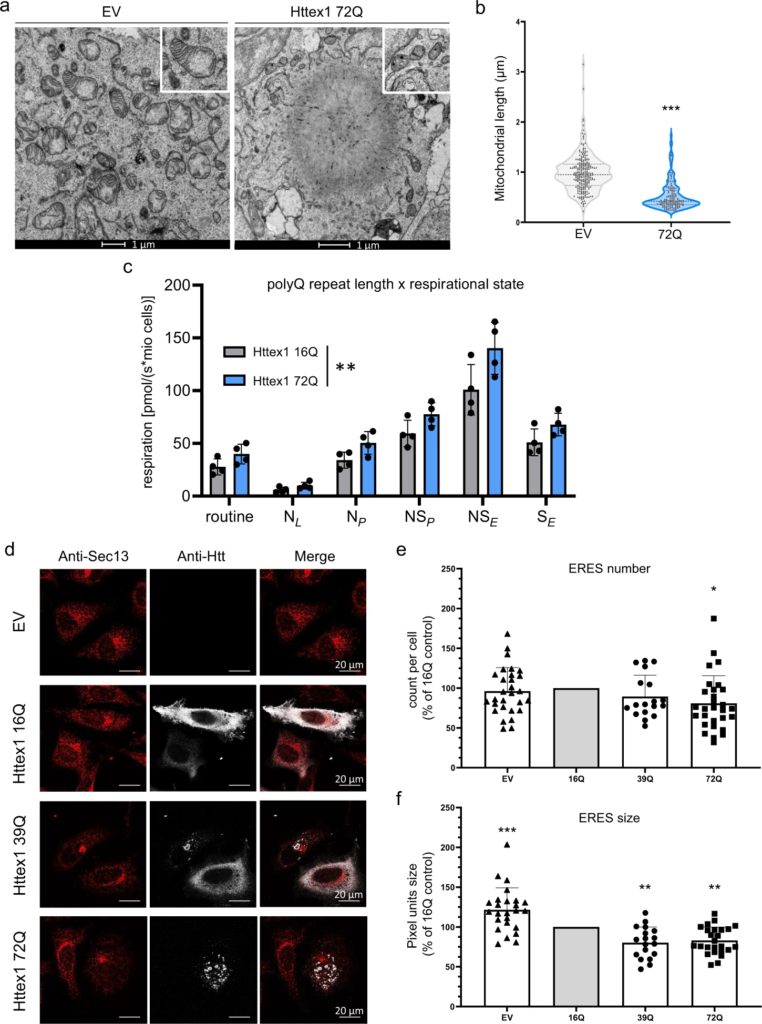
The remarkable accumulation of damaged mitochondria at the periphery of Httex1 72Q inclusions prompted us to investigate how Htt inclusion formation impacts mitochondrial functions. Quantification of mitochondrial length from EM-micrographs revealed a shorter mitochondrial profile associated with Httex1 72Q inclusions, as compared to HEK cells transfected with empty vector (Fig. 4a, b). Similar levels of the outer mitochondrial membrane protein VDAC1 suggest that this mitochondrial fragmentation was not associated with a decrease in mitochondrial density (Supplementary Fig. 13a). We hypothesized that the fragmentation and recruitment of mitochondria to Httex1 inclusions might be associated with respirational dysfunction. Therefore, we performed high-resolution respirometry on cells transfected with Httex1 16Q and 72Q for 48 h. Aggregates were only detected in mutant Httex1 72Q transfected cells (Supplementary Fig. 13b). We assessed different respirational states of mitochondria by high-resolution respirometry (Supplementary Fig. 13c-d). Httex1 72Q transfection resulted in significantly higher mitochondrial respiration than Httex1 16Q (Supplementary Fig. 4c). Together, our results demonstrate a clear impact of Httex1 72Q inclusion formation on mitochondrial morphology and function.
Formation of Httex1 72Q inclusions induces mitochondrial alterations and the reduction of ER-exit sites.
a Electron micrographs of mitochondria in HEK cells overexpressing empty vector (EV) or Httex1 72Q. The insets depict higher magnification of the mitochondria found at the periphery of the Httex1 72Q inclusions or in EV controls. Scale bars = 1 μm. b Measurement of the mitochondrial length with FIJI reveals a significant reduction in the size of the mitochondrial profile located in the proximity of the inclusions. An unpaired t-test was performed (two-sided) resulting in a p-value of 0.0006. An unpaired t-test was performed (two-sided) resulting in a p-value of 0.0006. c HEK cells from 4 independent experiments were gently detached for high-resolution respirometry (HRR) 48 h after transfection with indicated constructs. HRR was performed in respiration media. After the measurements of routine respiration, cells were permeabilized by digitonin, and different respirational states were subsequently induced using a substrate-uncoupler-inhibitor titration (SUIT) protocol. Routine respiration, NADH-driven, or complex 1-linked respiration after the addition of ADP (OXPHOS state) (NP), NADH- and succinate driven, or complex 1 and 2-linked respiration in the OXPHOS state (NSP), and in the uncoupled electron transport system (ETS) capacity (NSE), as well as succinate driven, or complex 2-linked respiration in the ETS state (SE) were assessed. The graphs represent the mean ± SD of 4 independent experiments. Two-way ANOVA showing a significant interaction between the respirational states and the polyQ repeat length (p-value = 0.004). d Representative confocal images of HeLa cells transfected with Httex1 16Q, 39Q, or 72Q or EV. Cells were fixed 48 h after transfection and immunostained. Httex1 was detected with the MAB5492 Htt antibody (grey), and ER exit sites (ERES) were detected with Sec13 (red). Scale bars = 20 μm. e, f ERES number (e) and size (f) quantifications from confocal imaging were performed using FIJI. The graphs represent the mean ± SD of 3 independent experiments represented as a relative percentage to Httex1 16Q control. One-way ANOVA followed by a Tukey honest significant difference [HSD] post hoc test was performed (p-values < 0.0001 for panels e and f). *P < 0.05, **P < 0.005, ***P < 0.001 for multiple comparisons.
Our EM analysis and 3D reconstruction further revealed the presence of ER cisternae at the periphery of the Httex1 inclusions. Stacked ER cisternae are usually formed by an increasing concentration of specific resident proteins or stress conditions92. The changes we observed in ER organization, together with the enrichment of proteins related to ER-Golgi trafficking inside the inclusions, prompted us to investigate whether ER functions were impaired upon the formation of the Httex1 inclusions.
A major function of the ER is the biogenesis of COPII carriers that ferry proteins and lipids to distal compartments. COPII carriers form at ER exit sites (ERES), which are ribosome-free domains of the rough ER. To determine whether the formation of Httex1 inclusions interferes with the homeostasis of the ERES, we used confocal imaging and quantified the number of ERES, labelled specifically by the COPII component Sec13 protein in HeLa cells expressing Httex1 72Q, Httex1 39Q, or Httex1 16Q (Fig. 4d). Our data show that the number of ERES was significantly reduced (~20%) only in cells containing Httex1 72Q inclusions (Fig. 4e). We next quantified the size of the ERES using the same imaging pipeline (Fig. 4f). Overexpression of Httex1 16Q caused a 20% reduction in the size of the ERES compared to the empty vector (EV). However, the reduction became much more significant in the cells carrying Httex1 39Q or Httex1 72Q, with a ~40% decrease compared to the empty vector and ~20% compared to Httex1 16Q. These results demonstrate that the formation of the Httex1 inclusions interferes with the formation and fusion of ERES. The remodeling of ERES in cells has been described primarily as an adaptive response to the protein synthesis level of ER with the number of ERES proportional to the cargo load. Interestingly, our proteomics results (Fig. 3a) showed that the TGF (Transforming Growth Factor) protein—which plays a central role in the biogenesis and organization of ERES—is sequestered in mutant Httex1 inclusions93. In line with our results, it has been previously shown that the depletion of TGF induced a dramatic reduction of the ERES sites94. Alternatively, the reduction of the ERES sites could represent early signs of cell vulnerability and toxicity induced by the presence of the Httex1 inclusions in cells.
Expression of Httex1 72Q in primary cortical neurons leads to the formation of dense and filamentous nuclear inclusions
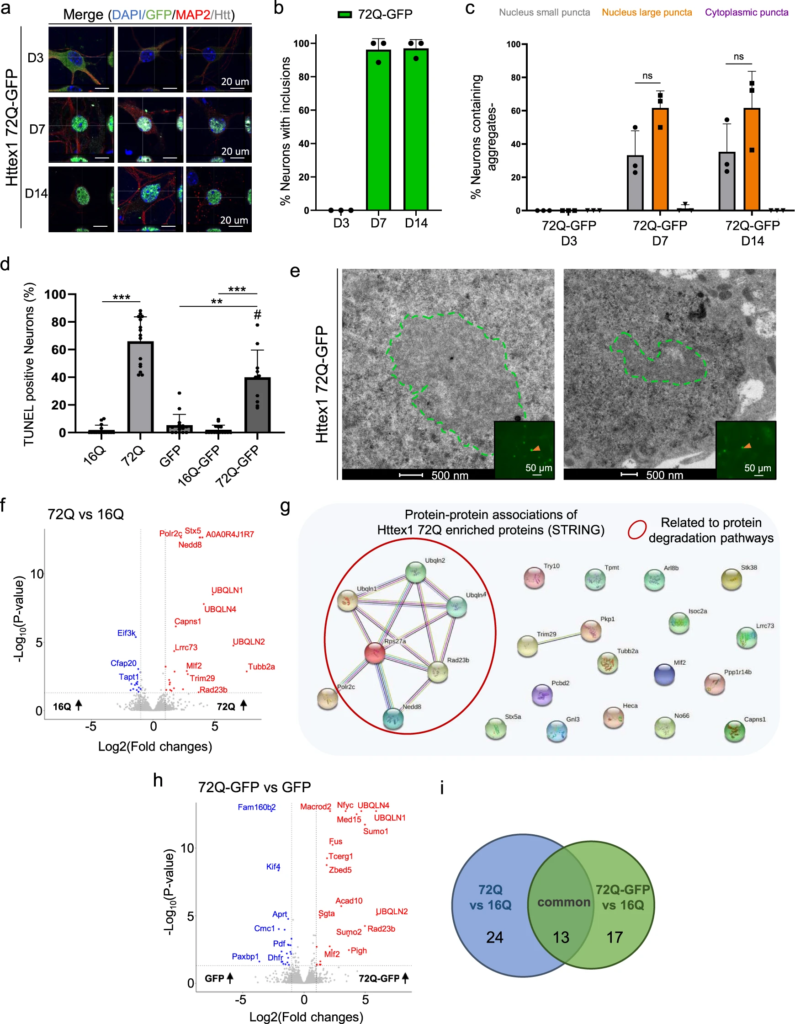
We next investigated the sequence determinants of Htt aggregation and inclusion formation in primary cortical neurons over time. As expected, none of the Httex1 16Q constructs (Httex1 16Q and ΔNt17 Httex1 16Q) overexpressed in neurons induced the formation of aggregates up to 14 days post-transduction (D14). Conversely, overexpression of Httex1 72Q induced the formation of round nuclear inclusions in almost 100% of transduced neurons, already at D3 (Fig. 5a, b, and S14). Less than 1% of the neurons showed cytoplasmic inclusions, either as puncta or with the ring-like morphology. Although no changes in the subcellular distribution of the inclusions were observed up to D14, we observed significant changes in the size and shape of the nuclear inclusions over time (Fig. 5c). At D3, the majority of the nuclear inclusions were detected as small (<1 μm) nuclear puncta (94%), and only a few appeared as large (~3–4 μm) inclusions (Fig. 5d). The ratio of small and large nuclear inclusions shifted slightly over time from 50:50 at D7 to 43:57 at D14 (Fig. 5d).
Confocal microscopy analysis and classification of the Httex1 inclusions formed in neurons revealed their morphological heterogeneity.
a Httex1 expression was detected by ICC staining combined with confocal imaging in primary cortical neurons, 3 (D3), 7 (D7), and 14 (D14) days after lentiviral transduction. Httex1 mutants were detected with the MAB5492 antibody. The nucleus was counterstained with DAPI (blue), and MAP2 was used to visualize the neurons (red). Scale bars = 20 μm. b Image-based quantification of the number of neurons containing Httex1 inclusions over time. The graphs represent the mean ± SD of 3 independent experiments. c Primary neurons were classified via the detection of Httex1 as diffuse or by the morphology of the detected Httex1 aggregates: (1) Small nuclear puncta; (2) at least one large nuclear inclusion, and (3) cytoplasmic inclusion. In addition, a subclass was created for neurons containing a large nuclear inclusion associated with nuclear condensation. Scale bar = 20 μm. d Image-based quantification and classification of the different morphologies of Httex1 inclusions based on the panel c. A minimum of n = 145 cells per condition were examined over 3 independent experiments. Data are presented as mean values + /− SD. Statistical analysis: One-way ANOVA followed by a Tukey [HSD] post hoc test was performed. p-values < 0.0001 for top and bottom panels and *P < 0.05, **P < 0.005, ***P < 0.001 for multiple comparisons. e Image-based quantification of neurons containing a large nuclear inclusion with a nuclear condensation. A minimum of n = 145 cells per condition were examined over 3 independent experiments. Data are presented as mean values + /− SD. Statistical analysis: Two-way ANOVA revealed no significant interaction (p-value = 0.0598) but a significant row factor samples (p-value = 0.0076) and column factor time (p-value = 0.0002) with *P < 0.05, **P < 0.005, ***P < 0.001 for multiple comparisons.
We next characterized the ultrastructural properties of the nuclear Httex1 72Q inclusions by CLEM. At D7, these inclusions appeared as dense and roughly round aggregates without distinctive core and shell structural organization (Fig. 6a and S15a). In these thin sections, the intranuclear Httex1 72Q inclusions appeared darker than the surrounding nucleoplasm and structurally different from the nucleolus (Fig. 6a and controls Supplementary Fig. 15c, d). The high density of these aggregates made it challenging to determine if they were made of filamentous structures. However, using electron tomography (ET), we were able to visualize and confirm the presence of filamentous structures inside these nuclear inclusions (Fig. 6band Supplementary Movie 3). The segmented filaments did not appear to be closely stacked in parallel but rather organized as a network of tortuous filaments. Neither EM nor ET imaging revealed the presence of membranous-, organelle- or vesicle-like structures inside or at the periphery of these inclusions. Although removal of the Nt17 domain (Fig. 6c) did not alter the ultrastructural properties or composition of the inclusions, it accelerated the formation of large aggregates significantly. As early as D3, ~60% of the neurons overexpressing ΔNt17 Httex1 72Q already contained large nuclear inclusions, compared to only ~6% for Httex1 72Q (Fig. 5d). At D14, almost all the aggregates formed in the ΔNt17 Httex1 72Q overexpressing neurons converted into the large nuclear inclusions (~90%) (Fig. 5d), compared to 57% for Httex1 72Q.
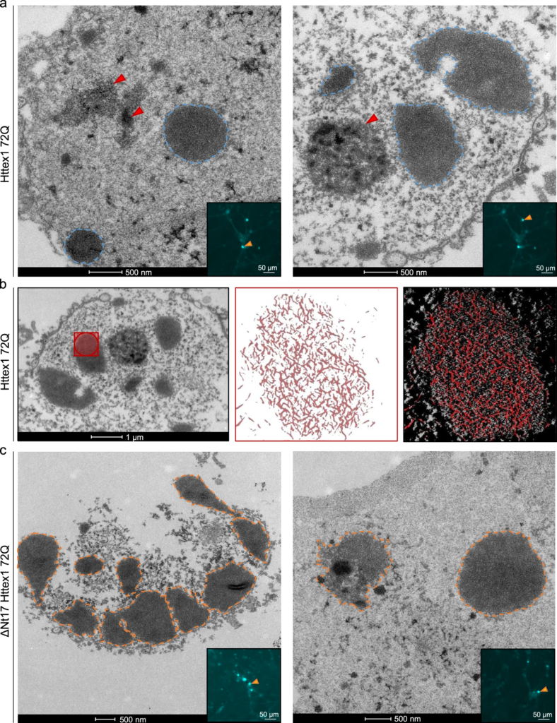
Ultrastructural analysis of the nuclear Httex1 72Q inclusions formed in primary cortical neurons shows granular and filamentous structures by CLEM and Tomography.
a Representative electron micrographs of Httex1 72Q inclusions formed 7 days after lentiviral transduction in mouse cortical primary neurons. Blue dashed lines represent the inclusions, and the red arrowheads the nucleolus. Scale bars = 500 nm for the EM images and 50 μm for the fluorescent image (inset). bElectron micrograph and corresponding tomogram (selected area in red) of a neuronal Httex1 72Q inclusion in panel a. Segmentation of the tomogram reveals the presence of a multitude of filaments. c Representative electron micrographs of ΔNt17 Httex1 72Q inclusions formed seven days after lentiviral transduction in mouse cortical primary neurons. Orange dashed lines represent the inclusions, and the red arrowheads the nucleolus. Scale bars = 500 nm for the EM images and 50 μm for the fluorescent image (inset).
Finally, among the small aggregates dispersed throughout the nucleus, a few were observed near the nuclear membrane, which often appeared damaged and ruptured (Figures S15a). Our data are in line with previous EM studies from human HD patients95 and HD mice models19,96showing nuclear ultrastructural changes, including altered nuclear membrane shape, nuclear invagination, and increased nuclear pore density in neurons bearing Httex1 inclusions. Consistent with these observations, the nuclei containing Httex1 72Q inclusions showed enhanced nuclear condensation over time (Fig. 5e), indicating increased neuronal toxicity, consistent with previous reports in HD patients97 and HD mice model96. Interestingly, despite the presence of the large inclusions as early as D3 in the neurons overexpressing ΔNt17 Httex1 72Q, we did not observe an earlier onset of cell death or a higher level of toxicity over time in these neurons compared to those overexpressing Httex1 72Q (Fig. 5e). This suggests a lack of correlation between cell death level and the size of the inclusions, with the large inclusions being less toxic.
These results demonstrate the formation of condensed mutant Httex1 fibrillar aggregates within intranuclear inclusions in neurons and suggest that the process of their formation and maturation is directly linked to neuronal dysfunctions and cell death.
The addition of GFP to the C-terminal part of Httex1 induces a differential structural organization and toxic properties of Httex1 inclusions
Given that the great majority of cellular models of HD rely on the use of fluorescently tagged Htt constructs, we next investigated the aggregation properties of mutant Httex1 fused to GFP (Httex1 72Q-GFP and Httex1 39Q-GFP). First, we assessed the morphology of Httex1 72Q-GFP cytoplasmic inclusions in HEK cells by ICC using a panel of Httex1 antibodies (Fig. 7a, band S16a). Confocal imaging revealed a diffuse GFP signal throughout the inclusions. Conversely, the Htt antibodies labeled mainly the outermost region of Httex1 72Q-GFP inclusions, but also faintly stained their centers (Fig. 7b). In contrast, the tag-free inclusions’ cores were not labeled by all the Htt antibodies tested. Thus, we hypothesized that the presence of the GFP tag results in the formation of less compact Httex1 inclusions. While Actin-F was found to colocalize with tag-free Httex1 inclusions [(Fig. 1b, Httex1 72Q), 39Q (Supplementary Fig. 9d, Httex1 39Q), and ΔNt17 Httex1 72Q (Supplementary Fig. 10a, ΔNt17 Httex1 72Q)], no specific enrichment of Actin-F was detected in the core or periphery of the Httex1 72Q-GFP inclusions. Furthermore, actin filaments were found exclusively at the periphery of the tag-free Httex1 72Q and 39Q (+/− ΔNt17) cytoplasmic inclusions (Fig. 1b and S10A). These results suggest a potential role of actin in the formation of Httex1 inclusion formation or maturation. Consistent with these observations, the actin cytoskeleton was one of the most dysregulated pathways according to a proteomic analysis conducted in human HD brains98, underscoring its importance in the development of HD pathology.
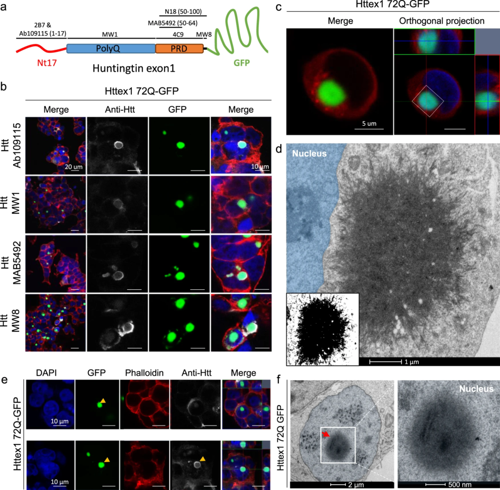
The addition of GFP to the C-terminal part of Httex1 induces a differential structural organization as revealed by ICC and CLEM.
a Epitope mapping of the Httex1 antibodies. b Httex1 72Q-GFP inclusions formed 48 h after transfection in HEK cells were detected by ICC staining combined with confocal imaging. All Htt antibodies showed strong immunoreactivity to the periphery of the Httex1 inclusions and modest immunoreactivity to the core. The nucleus was counterstained with DAPI (blue) and the F-actin with phalloidin (red). Scale bars = 20 μm (left-hand panels) and 10 μm (middle and right-hand panels). c, d 48 h post-transfection, HEK cells were fixed, and ICC against Httex1 was performed and imaged by confocal microscopy (c). Scale bars = 5 μm. The selected area of the cells (white square) was then examined by EM (d). A binary image (inset) was generated from the electron micrograph using a median filtering and Otsu intensity threshold, allowing for a better distinction of the inclusions’ morphology. Scale bars = 500 nm. e Representative confocal images of Httex1 72Q-GFP nuclear inclusions formed in HEK cells 48 h after transfection. Httex1 expression (grey) was detected using a specific antibody against the N-terminal part of Htt (amino acids 1-17; 2B7 or Ab109115), and GFP (green) directly visualized in the appropriate channel. The nucleus was counterstained with DAPI (blue), and phalloidin (red) was used to stain filamentous actin. Httex1 nuclear inclusions are indicated by yellow arrowheads. Scale bars = 10 μm. f Electron micrograph of a representative Httex1 72Q-GFP inclusion. The white square indicates the area shown at higher magnification in the right-hand panel. The nucleus is highlighted in blue. Scale bars = 2 μm (left-hand panel) and 500 nm (right-hand panel).
Next, we performed a more in-depth analysis of the Httex1 72Q-GFP cellular inclusions formed in HEK by CLEM (Fig. 7c, d). The Httex1 72Q-GFP inclusions were organized as a highly dense network of fibrils, which were more homogenously stained (Fig. 7d and S16c) and did not exhibit the core and shell architecture that is characteristic of the tag-free Httex1 72Q inclusion (Fig. 1d and S16d). Closer examination of the inclusions showed that they were composed of densely packed fibrils that exhibited a striking resemblance to the fibrillar aggregates formed by mutant Httex1 proteins in a cell-free system (Fig. 7d and S16b). Structural analysis of Httex1 72Q-GFP by high-pressure freezing fixation (Supplementary Fig. 4b) also revealed densely packed fibrils radiating from the inclusions. The center of the inclusions was not well resolved, but thicker fibrils could clearly be observed radiating at the periphery with increased spacing between them compared to tag-free Httex1 72 inclusion (Supplementary Fig. 4a). A portion of Httex1 72Q-GFP inclusions exhibited perinuclear localization. In some cases, the accumulation of fibrils near the nuclear membrane leads to apparent distortion of the nucleus but without membrane disruption (Fig. 7d and S17a). Overall, we observed no significant differences in diameter and distance from the nucleus for all the Httex1 (+/− GFP) inclusions imaged in HEK cells (Supplementary Fig. 18).
Next, we investigated the ultrastructure properties of Httex1 72Q-GFP inclusions present in the nucleus of the HEK cells (Fig. 7e, f). No significant differences in terms of the organization were observed between nuclear and cytoplasmic Httex1 72Q-GFP inclusions by both GFP detection and antibody staining (Fig. 7e). The Httex1 72Q-GFP nuclear inclusions were also enriched in fibrillar structures. The 3D reconstruction generated from the series of electron micrographs revealed much fewer membranous structures in both Httex1 39Q-GFP and Httex1 72Q-GFP inclusions (Supplementary Fig. 19) compared to tag-free Httex1 inclusions (Fig. 1f, h). Moreover, consistent with EM observations, no neutral lipids were found in Httex1 39Q-GFP and Httex1 72Q-GFP inclusions (Supplementary Fig. 10b-c). Interestingly, neither the length of the polyQ repeat nor the presence or removal of the Nt17 domain seem to significantly alter the size, morphology, or structural properties of the inclusions formed by mutant Httex1 proteins fused to GFP (Supplementary Fig. 20). This is different from what we observed for cells expressing tag-free Httex1, in which the increase of the polyQ length led to inclusions with distinct morphologies and organizational features. These findings suggest that the addition of GFP significantly alters the mechanism of Httex1 aggregation and inclusion formation.
To obtain an even clearer picture of these differences, we next performed EM under detergent-free conditions to preserve the internal membranes and structures of the inclusions. We observed that the length of the polyQ repeat did not influence the size of the Httex1 39Q-GFP and 72Q-GFP inclusions (Supplementary Fig. 18b) or their overall architecture in HEK cells. The Httex1 39Q-GFP inclusions are composed of radiating fibrils and are thus similar to the Httex1 72Q-GFP inclusions, although slightly less dense. The 3D reconstruction of the inclusions showed the presence of ER and mitochondria in their periphery, but few membranous structures were internalized (Supplementary Fig. 19b yellow arrowhead) as compared to tag-free Httex1 39Q and Httex1 72Q inclusions. A similar analysis of Httex1 ΔNt17 39Q-GFP and Httex1 ΔNt17 72Q-GFP cellular inclusions (Fig. S20a–d) also revealed no effect of Nt17 deletion on the ultrastructure of the inclusions or their interactions with the surrounding organelles. These results confirm that mutant Httex1 aggregation and inclusion formation mechanisms are significantly altered by the addition of the GFP.
We also assessed how the presence of the GFP tag might influence the kinetics of aggregation, the morphology, subcellular localization, and toxicity of the nuclear inclusions in primary cortical neurons (Fig. 8 and S21). First, we observed that the presence of the GFP tag slows down the aggregation rate of Httex1 72Q in contrast to the tag-free Httex1 72Q, the aggregation was significantly delayed, as evidenced by the absence of nuclear or cytoplasmic aggregates or inclusions at D3 (Fig. 8a, b). However, at D7, almost all the Httex1 72Q-GFP proteins appeared in the form of small nuclear puncta (~35%) or large nuclear inclusions (~60%) (Fig. 8c). The proportion of small puncta vs. large inclusions did not change over time, up to D14 (Fig. 8c). Interestingly, the subcellular localization, morphology, and size distribution of the inclusions observed by confocal imaging were not impacted by the presence of the GFP tag. However, the Httex1 72Q-GFP aggregates exhibited a significantly reduced toxicity compared to tag-free Httex1 72Q in neurons (Supplementary Fig. 21d). Even at D14, chromatin condensation did not exceed 10% in these neurons (Supplementary Fig. 21d), whereas ~50% of the neurons overexpressing the tag-free Httex1 72Q construct were already dead (Fig. 5e). In addition, the TUNEL cell death assay revealed a dramatic increase (~60%) in DNA fragmentation in the cortical neurons bearing the intranuclear Httex1 72Q inclusions compared to only 40% in neurons expressing Httex1 72Q-GFP (Fig. 8d).
The GFP tag alters the kinetic, ultrastructural properties, protein content and toxicity of Httex1 72Q inclusions in neurons.
a Httex1 72Q-GFP was directly visualized with GFP (green) at 3 (D3), 7 (D7), and 14 (D14) days after lentiviral transduction. The nucleus was counterstained with DAPI, and neurons were stained with the MAP2 antibody. Scale bar = 20 μm. b Image-based quantification of the number of transduced neurons bearing Httex1 72Q-GFP inclusions over time. c Morphological and subcellular localization classifications (see Fig. 5c) of the 72Q-GFP inclusions formed in transduced neurons. n = 219 cells were examined over 3 independent experiments. d TUNEL cell death assay in cortical neurons transduced with Httex1. b, c, d The graphs represent the mean ± SD of 3 independent experiments. b, d One-way ANOVA followed by an HSD post hoc test was performed. *P < 0.05, **P< 0.005, ***P < 0.001 for multiple comparisons. #P < 0.05 (72Q vs. 72Q-GFP). c Two-way repeated-measures ANOVA revealed no significant differences. eRepresentative EM images of Httex1 72Q-GFP inclusions formed in cortical neurons at D7 post-transduction. Green dashed lines represent Httex1 inclusions. Scale bars = 500 nm and 50 μm (inset). f-i Urea soluble proteins extracted from cortical neurons expressing Httex1 72Q or Httex1 16Q (f) or Httex1 72Q-GFP or GFP (h) at D7 post-transduction were analyzed using LC-MS/MS. n = 3 independent experiments. Identified proteins were plotted using volcano plots (f, Httex1 72Q vs. Httex1 16Q; h, Httex1 72Q-GFP vs. GFP). Dotted lines represent the false discovery rate <0.05, and an absolute log2 fold change threshold of significance of 1 was assigned. Upregulated proteins are represented in red, downregulated in blue and non-significant proteins in grey. g The online platform STRING underscores an association between the upregulated Httex1 72Q proteins and protein related to the degradation pathways (red circle). i Venn diagram comparison of proteins enriched in Httex1 72Q vs. Httex1 16Q (blue) to Httex1 72Q-GFP vs. Httex1 16Q (green). 24 proteins were unique to Httex1 72Q vs. Httex1 16Q (44.4%), 17 unique to Httex1 72Q-GFP vs. Httex1 16Q (31.5%) and 13 (24.1%) common to both co-aggregated proteins. Source data are provided as a Source Data file.
Next, we investigated how the presence of the GFP tag might impact the ultrastructural properties of these neuronal inclusions (Fig. 8e and S22). The nuclear inclusions formed in the presence of the GFP tag exhibited a round shape but displayed a less dark staining density compared to the inclusions formed by the untagged Httex1 72Q protein (Fig. 6a). Due to their lower density, the presence of filamentous structures organized as an entangled arrangement throughout the GFP tagged inclusion could be detected. Finally, these inclusions were localized throughout the nucleus but always distant from the nuclear membrane (Supplementary Fig. 22).
Altogether, our results demonstrate that the addition of the GFP significantly slows the initiation of mutant Httex1 fibrillization and inclusion formation in the nucleus but accelerates the maturation of the aggregates once formed, resulting in predominantly large inclusions with reduced toxicity in neurons. The lack of physical interaction between the Httex1 72Q-GFP inclusions and the nuclear envelope and/or the absence of small puncta aggregates in neurons expressing Httex1-72Q-GFP could explain their reduced toxicity (Fig. 8d, e).
The Httex1 72Q and Httex1 72Q-GFP nuclear and cytoplasmic inclusions exhibit distinct proteome composition
To gain further insight into the biochemical composition of the Htt nuclear inclusions, the molecular interactions and mechanisms driving their formation and maturation, we next investigated the proteome content of nuclear inclusions formed in primary neurons (Supplementary Fig. 23). Proteomic analysis showed that 23 proteins were significantly enriched in the Httex1 72Q inclusions compared to Httex1 16Q (Fig. 8f). These proteins were analyzed by protein-protein association using the online platform STRING (Fig. 8g) and classified using GO term analysis by Cellular component, Molecular function, and Biological process (Supplementary Fig. 24). The proteins identified include the transcription factors PCBD2 and Mlf2, the RNA/DNA binding protein (GNL3), DNA-dependent RNA polymerase (Polr2c), histone lysine demethylase (No66), DNA genome nucleotide excision repair (Rad23B), and proteins linked to chromosome segregation (ARL8B and Tubb2a). In line with our results, ARL8B, Tubb2A, Polr2 sub-units, Mlf2, GNL3, and Rad23B proteins were also found enriched in the insoluble fractions of Q175 HD mice brains99 and Tubb2a in R6/2 mice100 nuclear inclusions. Interestingly, the gene expression level of these nuclear proteins was also significantly increased in HD post-mortem brain (PCBD2)101 and symptomatic HD patients (Mlf2, GNL3, Polr2, and ARL8B)102. Altogether, this suggests that the loss of key nuclear proteins due to their sequestration by the pathological Htt inclusion is compensated in neurons by increasing gene expression levels. However, this seems insufficient to compensate for the loss of their biological functions, as reflected by the high level of nuclear alterations observed in our neuronal HD model, including chromatin condensation, nuclear fragmentation, and nuclear envelope integrity loss (Fig. 6).
In addition to the nuclear proteins, STRING analysis (Fig. 8g), together with GO term classification of cellular components (Supplementary Fig. 24a), the molecular functions (Supplementary Fig. 24B), and the biological process annotations (Supplementary Fig. 24c) revealed the enrichment of a cluster of proteins [Ubiquilin (ubqln) 1, 2 and 4; Rps27a, Rad23B and Nedd8, red circle, Fig. 8g] related to the protein degradation machinery, including the UPS, autophagy and ERAD pathways. In line with our findings, all these proteins were also found enriched in the insoluble fractions of the Q175 HD mice brains, while the Ubiquilin family members (Ubqln 1, 2, or 4)103,104,105 were also shown to be sequestered in R6/2 mice nuclear inclusions100 and in the Httex1 nuclear inclusions formed in the PC12 neuronal rat precursor106. Interestingly, ubiquilin proteins have been previously shown to translocate from the cytoplasm to the nucleus or have an upregulated expression level in the nucleoplasm concomitantly with the early stage of the formation of the neurofibrillary tangles in AD post-mortem brain tissues107. Furthermore, Nedd8, originally shown to colocalize with ubiquitin and the proteasome components in the cytoplasmic inclusions108,109 found in several neurodegenerative disorders, has recently been shown to promote nuclear protein aggregation as a defense mechanism against proteotoxicity110. Moreover, our analysis revealed 5 proteins known as Htt interactors (based on the HDinHD database) among the proteins significantly different between Httex1 72Q compared to Httex1 16Q in primary neurons (Supplementary Data 1).
Finally, we also assessed how the presence of the GFP tag might influence the protein content of the neuronal intranuclear inclusions (Fig. 8h). Similar to the Httex1 72Q aggregates, ~45% of the proteins enriched in the insoluble fraction of the Httex1 72Q-GFP aggregates were related to nuclear biological processes and functions, including transcription factors (Mlf2106, nfyc-1, TCERG1, Med15), DNA-chromatin binding proteins (ZBED5, N6AMT1, and Actl6b), DNA genome nucleotide excision repair protein (Rad23B), and the DNA/RNA-binding protein FUS. In addition, Httex1 72Q-GFP aggregates were also enriched in proteins from the promyelocytic leukemia nuclear bodies (Sumo 1 and 2) known as nuclear membraneless compartments involved in genome maintenance such as DNA repair, DNA damage response, telomere homeostasis, and which is also associated with apoptosis signaling pathways (Supplementary Fig. 25). Furthermore, proteins from the ubiquitin-proteasome system (Bag5, Fus, Rad23b, Sgta, Sumo1, Sumo2, Ubqln1, Ubqln2, Ubqln4), the ERAD (Sgta, Ubqln1, Ubqln2), autophagy (Ubqln1, Ubqln2, Ubqln4), and sumoylation (Sumo1, Sumo2) pathways were also highly enriched in the insoluble fraction of the Httex1 72Q-GFP aggregates (Supplementary Fig. 25a, b). Interestingly, the proteome of the Httex1 72Q-GFP aggregates formed in our HD neuronal model shared 40% of the proteins also found enriched in the insoluble fraction of Httex1 74Q-GFP nuclear inclusions formed in the PC12 neuronal rat precursor cell line106. This highlights a strong similarity between the pathways and proteins involved in the aggregation of Httex1 and inclusion formation in rodent neuronal cells.
We next compared the proteins identified between tag-free Httex1 72Q inclusions and Httex1 72Q-GFP (Fig. 8i). 24 proteins were unique to Httex1 72Q (44.4%), 17 unique to Httex1 72Q-GFP (31.5%) and only 13 (24.1%) common to both type of inclusions. Most of the unique proteins found in the tag-free or GFP-tagged Httex1 72Q inclusions were related to nuclear functions, the ubiquitin-proteasome system, ERAD, and autophagy pathways. Moreover, among the 13 common proteins, most were linked to the protein degradation machinery (i.e., ubiquilins 1, 2, and 4, and the Rad23b and Nedd8 proteins).
Although the tag-free Httex1 72Q and Httex1 72Q-GFP inclusions share several proteins related to the degradation machinery, more than 75% of the co-aggregating proteins are different, which could explain the drastic differences in neuronal toxicity associated with each type of these inclusions.
As the majority of the inclusions formed in neurons are nuclear, we used HEK cells to further assess how the GFP tag influences the proteome of the cytoplasmic Httex1 inclusions (Supplementary Fig. 11a). Volcano plot analysis showed 492 proteins significantly enriched in the insoluble fraction of the HEK cells overexpressing Httex1 72Q-GFP as compared to those expressing GFP (Fig. 9a). We observed a significant enrichment of the endogenous HTT protein, suggesting that, as for the tag-free Httex1 72Q, mutant Httex1-GFP inclusions can recruit the endogenous HTT protein (Supplementary Fig. 11e).
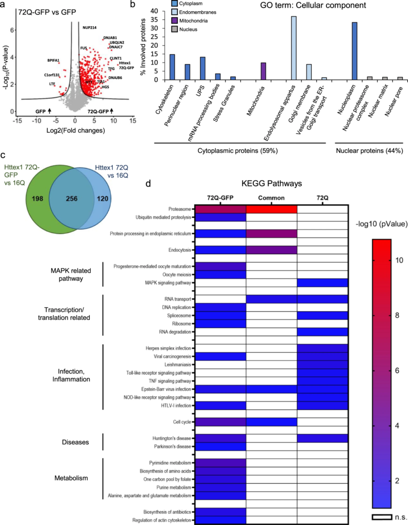
Proteomic analysis of Httex1 72Q-GFP in the Urea soluble fraction revealed 55% differences compared to tag-free Httex1 72Q for the proteins co-aggregated with Httex1 in HEK cells.
a Identified proteins were illustrated using a volcano plot for the comparison of protein levels identified in the Urea soluble fraction 48 h after Httex1 72Q-GFP or GFP transfection in HEK cells, in 3 independent experiments. Mean difference (Log2 (Fold-Change) on the X-axis) between the Urea soluble fraction of HEK cells overexpressing Httex1 72Q-GFP or GFP were plotted against significance (Log10 (p-Value) on the Y-axis (T-Test)). A false discovery rate (FDR) of 0.05 and a threshold of significance S0 = 0.5 were assigned for the subsequent analyses. b Cell compartment classification of the proteins significantly enriched in the Urea soluble fraction of the HEK cells overexpressing Httex1 72Q-GFP versus those expressing GFP. Gene Ontology (GO) enrichment analyses were determined by DAVID analysis (Significant for p values < 0.05 adjusted for multiple testing based on Benjamini and Hochberg). c To compare the results obtained with Httex1 72Q-GFP to tag-free Httex1 72Q, we represented the proteins enriched in Httex1 72Q vs. Httex1 16Q compared to the proteins enriched in Httex1 72Q-GFP vs. Httex1 16Q using a Venn diagram. In total, 256 proteins (44.6%) were found similar between the two conditions, 198 proteins (34.5%) were unique for Httex1 72-GFP enrichment, and 120 proteins (20.9%) were for Httex1 72Q enrichment. d Kyoto Encyclopedia of Genes and Genomes (KEGG) Pathway Analysis of co-aggregated proteins with Httex1 72Q and Httex1 72Q-GFP. The heat map represents significant pathways (KEGG) enriched in the Urea soluble fractions extracted from the Venn diagram (c): co-aggregated proteins unique to Httex1 72Q, unique to Httex1 72Q-GFP, and common to both conditions, all compared to Httex1 16Q control. Source data are provided as a Source Data file.
The cytoplasmic components enriched in the insoluble fraction of Httex1 72Q-GFP were similar to those found with tag-free Httex1-72Q and were part of the endomembrane system (~50%, light blue Fig. 9b), the cytoskeleton, the perinuclear region, the UPS, mRNA processing bodies, and stress granules (Fig. 9b, dark blue). However, in contrast to the tag-free Httex1 72Q insoluble fractions, in which no mitochondrial protein was found to be enriched, 10% of the proteins enriched in the Httex1 72Q-GFP insoluble fraction were related to the mitochondria compartment (purple Fig. 9b). The nuclear proteins found in the Httex1 72Q-GFP insoluble fraction belong to similar nuclear compartments as those identified previously in the Httex1 72Q insoluble fraction (Fig. 9b, grey).
Biological process (Supplementary Fig. 26) and canonical pathway (Supplementary Fig. 27) analyses revealed that the UPS and chaperone machinery were the most enriched terms, as previously observed in the tag-free insoluble fraction. This indicates that the process of aggregation of Httex1 itself, regardless of the presence of the GFP tag, leads to the sequestration of the key cellular machinery responsible for protein folding and protein degradation. This could compromise the cell’s ability to prevent the accumulation or clearance of Httex1 aggregates. Although in different proportions, most of the biological processes and pathways significantly enriched in the tag-free Httex1 72Q insoluble fraction, such as inflammation, transcription, HD signaling, and cell death, were also detected in the Httex1 72Q-GFP insoluble fraction.
Having identified the proteins significantly enriched in both tag-free and GFP-tag 72Q inclusions, we next determined which proteins were unique to each type of inclusion. Toward this goal, we used a Venn diagram to compare the lists of proteins significantly enriched in our volcano plot analyses [(Httex1 72Q vs. Httex1 16Q insoluble fractions) vs. (Httex1 72Q-GFP vs. Httex1 16Q insoluble fractions)] (Fig. 3a and S11c). Figure 9c shows that ~45% (256 proteins) were found in both the tag-free and the GFP-tag Httex1 72Q inclusions. In total, 120 proteins (20.9%) were unique to the Httex1 72Q insoluble fraction, and 198 proteins (34.5%) were unique to the Httex1 72Q-GFP. Overall, we found 55% different proteins among the proteins that co-aggregate with or are sequestered in Httex1 72Q vs. 72Q-GFP inclusions, both compared to Httex1 16Q. We used the Kyoto Encyclopedia of Genes and Genomes (KEGG) pathway analysis to classify the list of proteins in Httex1 inclusions (Fig. 9d). The proteasome, protein processing in the endoplasmic reticulum, and endocytosis were among the most enriched pathways from the common co-aggregated proteins. Those 3 terms were also enriched significantly for Httex1 72Q-GFP, indicating the involvement of other unique proteins enriched in those pathways compared to Httex1 72Q. The KEGG classification revealed two distinct clusters for Httex1 72Q and Httex1 72Q-GFP, as well as additional differences (Fig. 9d). Proteins related to infection and inflammatory pathways (e.g., Herpes simplex infection, TNF signaling, and Toll-like receptor signaling) were found to be unique to Httex1 72Q, whereas proteins related to metabolism were specifically enriched for Httex1 72Q-GFP. Our proteomic analysis highlights that the addition of a fluorescent tag such as GFP significantly alters not only the mechanism of Htt inclusion formation but also the Htt interactome and thus the biochemical composition of inclusions.
Httex1 72Q inclusions induce higher respirational dysfunction and a stronger decrease of ERES number compared to Httex1 72Q-GFP in HEK cells
Next, we investigated how the aggregation of mutant Httex1-GFP and the formation of cytoplasmic inclusions affect the mitochondrial and ER-related functions. First, we measured the mitochondrial respiration in different respirational states in HEK cells (Supplementary Fig. 13c, d). Compared to our findings with the tag-free mutant Httex1 72Q (Fig. 4c), the increased mitochondrial respiration was significant but less pronounced for Httex1 72Q-GFP than for Httex1 16Q-GFP (Supplementary Fig. 13e). Given the role of mitochondria as a major source of reactive oxygen species (ROS), with implications of ROS in both neurodegenerative disease and cellular protective signaling cascades111,112, we suspected that mitochondrial ROS production might be affected by the mitochondrial fragmentation observed in regions close to tag-free Httex1 72Q and absent for Httex1 72Q-GFP (Supplementary Fig. 28a, b). To test this hypothesis, we used an amplex red assay to measure mitochondrial ROS (superoxide and hydrogen peroxide) production concurrent with mitochondrial respiration (Supplementary Fig. 13d). We observed no significant differences in mitochondrial ROS production between the tag-free or GFP tagged Httex1 72Q or 16Q (Supplementary Fig. 28c).
Mitochondrial fragmentation113 and inflammation114 are hallmarks of Htt-induced neurodegeneration. Recent studies reported a major role of mitochondrial fission and fusion homeostasis in HD and also revealed specific mitochondrial fragmentation induced by mutant Htt using electron microscopy in STHdh neurons115,116. Interestingly, only tag-free Httex1 72Q overexpression results in mitochondrial fragmentation and is characterized by the strong accumulation of inflammation-linked proteins in the aggregates. We speculate that the observed hyper-activation of mitochondrial respiration (in the absence of significantly increased ROS production) is an adaptation of the inclusion-bearing cells in order to generate sufficient energy by oxidative phosphorylation for increased energetic demands for unfolding or clearance of aggregating proteins. A similar hyperactivation of mitochondrial respiration has recently been reported for alpha-synuclein aggregates117. These authors hypothesized that the hyper-respiration might represent a pathogenic upstream event to alpha-synuclein pathology. We think that this may also be the case for untagged Httex1 72Q induced pathology. However, based on our extensive proteomic analyses of the inclusions formed by Httex1 72Q or Httex1 72Q-GFP and comparing the functionality of organelles in our cellular model, we can provide a more elaborate hypothesis. In the tag-free Httex1 72Q condition, mitochondrial hyperactivity coincided with both mitochondrial fragmentation and the detection of more inflammation-related proteins in the aggregates as compared to the GFP condition. We speculate that mitochondrial fragmentation initially may be protective, as it enhances mitochondrial mobility necessary for cell repair processes118. Upregulation of mitochondrial respiration would be a reasonable consequence of elevated ATP requirements for mitochondrial and protein transport related to aggregation formation and proteostatic processes including ATP-requiring chaperone-, proteasome and autophagy processes.
Finally, we also investigated whether Httex1-GFP inclusions interfere with the homeostasis of ERES by comparing the size and the number of ERES in HeLa cells overexpressing Httex1 16Q-GFP to those expressing mutants GFP-tagged Httex1 39Q or 72Q (Supplementary Fig. 29a). We found that cells containing Httex1 72Q-GFP inclusions caused a decrease in ERES number (Supplementary Fig. 29b), although non-significant, compared to Httex1 72Q, which showed a 20% reduction (Fig. 4e). The reduction of ERES size was significant for cells expressing Httex1 72Q-GFP but not Httex1 39Q-GFP compared to Httex1 16Q-GFP (Supplementary Fig. 29c). Thus, the effect of Httex1-GFP inclusions on ERES is present but is less pronounced compared to that observed in cells containing tag-free Httex1 inclusions. These observations are consistent with recent cryo-ET studies suggesting decreased ER dynamics near Httex1-GFP inclusions12.
Overall, our results showed that the cellular organelle responses and adaptation to inclusion formation were different for the Httex1 72Q-GFP inclusions than for the Httex1 72Q inclusions, consistent with the GFP-dependent changes observed in the proteome and the ultrastructural properties of Httex1 cytoplasmic inclusions.
Discussion
Pathological inclusions in neurodegenerative diseases have been shown to have a complex organization and composition. For example, it has been recently demonstrated that Lewy bodies (LB) isolated from Parkinson’s disease brains or LB-like inclusions in primary neurons are composed of not only filamentous and aggregated forms of alpha-synuclein but also a complex milieu of lipids, cytoskeletal proteins, and other proteins and membranous organelles, including mitochondria and autophagosomes119,120,121,122. Studies in neuronal cultures also showed that the recruitment of lipids and membranous organelles during LB formation and maturation contribute to organelles’ dysfunctions and lead to synaptic dysfunction and neurodegeneration. In line with these findings, our EM data, together with 3D reconstructions, revealed the presence of membrane fragments and vesicles entrapped in the core of the Httex1 inclusions. Consistent with these observations, our proteomic analysis revealed that 24% of the proteins enriched in the inclusions fraction belong to the endolysosomal compartments, the Golgi apparatus, and the trans-Golgi network (Fig. 3). In addition, mitochondria and ER were found in the periphery of inclusions, as previously reported in cellular models15 and human tissue95,123. We hypothesized that sequestration of key functional proteins, together with lipids, endomembranes, and organelles inside the Httex1 inclusions, could challenge cellular homeostasis. In line with this hypothesis, our electron micrographs revealed that the mitochondria associated with Httex1 72Q inclusions were fragmented and often exhibited disorganized or depleted cristae (Fig. 4a). These changes in mitochondrial morphology were associated with dysregulation of the mitochondrial respiration (Fig. 4c), consistent with previous studies demonstrating that mutant Htt aggregates interact directly with outer mitochondrial membranes (in STHdh cells)124 and induce mitochondrial fragmentation (in primary neurons)125. Defects in mitochondrial respiration have also been observed in HD patients’ brains, especially defects of complexes II and IV of the respiratory chain may impair oxidative phosphorylation126,127,128.
The ER at the periphery of the Httex1 72Q inclusions were also affected at the structural level, as shown by their morphological reorganization in rosette or “stacked cisternae”, and at the functional level, as evidenced by the dysregulation of the ERES homeostasis (Fig. 4d-f). The formation of ER rosettes indicates the accumulation of proteins in the smooth ER92 and is thought to result from low-affinity binding and export defects, which can be caused by unfolded Htt proteins but is not necessarily linked to ER stress. Kegel and colleagues129 also reported the presence of ER cisternae next to aggregates formed by the expression of a large Htt fragment fused to a FLAG tag in clonal striatal cells. The formation of Httex1 inclusions seems to drive the reorganization of the ER network in their periphery but did not lead to ER protein sequestration, as shown by the quantitative proteomic analysis, which did not reveal significant ER proteins trapped inside the inclusions. The presence of ribosomes and ER membrane deformation was previously detected close to the periphery of Httex1 inclusions by cryo-ET12 and was linked to a strong reduction in ER dynamics. In the same study12, the periphery of the inclusions was immunoreactive to several components of the ER-associated degradation (ERAD) machinery (e.g., Erlin-2, Sel1L). These observations are consistent with our ICC data showing the presence of ER chaperone BIP at the outer periphery but not inside the inclusions (Supplementary Fig. 5). We also observed ERES modulation, which can be explained by the sequestration of specific proteins required for their fusion130. Consistent with these observations, our proteomic analysis showed the enrichment of proteins specific to the ER-Golgi trafficking. ERES modulation can also be explained by a reduction of the biosynthetic capacity of this compartment, as ERES have been shown to adapt to the amount of secretory burden to which they are exposed131. The recruitment and perturbation of the ER network might contribute to cytotoxicity during inclusion formation, but on its own does not appear to be sufficient to cause overt toxicity. Consistent with this hypothesis, the ERAD and Ca2+ signaling pathways have been extensively reported to be dysregulated in various cellular and animal models using different Htt fragments132,133,134,135,136,137. Overall, we showed that cytoplasmic Httex1 inclusion formation and maturation involves a complex interplay between Httex1 aggregates and organelles.
Although previous studies have suggested the formation of ring-like nuclear inclusions, this architecture was defined by the peripheral staining of nuclear proteins and not by the peripheral staining of Htt as observed in the case of cytoplasmic inclusions (Fig. 1). Our in-depth characterization of the Httex1 72Q inclusions at the ultrastructure level revealed distinct differences in the organization and architecture between the cytoplasmic and nuclear Htt inclusions. The cytoplasmic Httex1 72Q inclusions exhibit a dense core and shell morphology and are composed of highly organized fibrillar aggregates in both the core and the periphery. In contrast, nuclear Htt inclusions, in both HEK cells or primary cortical neurons, exhibited less complex ultrastructural properties, appeared as homogenous structures without the core and periphery morphology and were devoid of endomembranes or vesicles (Figs. 2 and 6a). These observations are consistent with most of the Htt nuclear inclusions found in HD mice models, which do not display the core and shell organization19,32.
Our new data on nuclear neuronal Htt inclusion revealed that tag-free mutant Httex1 expression in primary neurons leads to the formation of granulo-filamentous nuclear inclusions, similar to previous ultrastructural studies performed in HD patients brains and in vivo models of HD, including R6/2 mice19,32 and transgenic rats138. However, in most of these studies, the ultrastructural properties of the filaments were less evident compared to the well-resolved filamentous structure we and others12 showed using electron tomography. For example, Tagawa and colleagues also observed nuclear inclusions of tag-free Httex1, but their data did not reveal the level of ultrastructural details and insights that our study provides139.
Our results also suggest that the intracellular environment and interactions with lipids and endomembranes or vesicles are key determinants of the structural and molecular complexity of the inclusions and that different mechanisms drive the formation and maturation of the nuclear and cytoplasmic inclusions. Such differential characteristics between cytoplasmic and nuclear inclusions might account for the differential cellular dysfunction and toxicity associated with these two types of Htt inclusions29,140,141,142,143. Furthermore, the differential toxicity seen for Htt nuclear inclusions in primary neurons compared to HEK 293 cells indicate a cell-type dependent Htt toxicity that might arise from distinct Htt mechanisms of aggregation or interaction with cellular proteins or differences in the resilience of the different cells based for example on distinct oxidative stress tolerance and immune functions/inflammation.
We speculate that the formation of the core of the inclusions is guided predominantly by intermolecular interactions involving the polyQ domain and could be initiated by rapid events that are potentially driven by phase separation, as recently described by several groups14,39,144,145. The rapid formation of the aggregate’s core does not allow for the regulated recruitment of other proteins and organelles (Figs. 1, 2, and 10a), as evidenced by the fact that most antibodies against proteins found in Htt inclusion stain the periphery rather than the core of the inclusions48. The Nt17 domain most likely plays key roles in the initial oligomerization events and, possibly, the packing or lateral association of the fibrils. Once this core of dense fibrils is formed, the fibrils at the periphery continue to grow through the recruitment of endogenous soluble proteins (Figs. 3 and 10a). Because this growth phase is slower, it allows the fibrils to interact with and/or recruit other proteins into the inclusions. Consistent with this hypothesis, Matsumoto and colleagues showed that the transcription factors TATA-binding protein (TBP) and CREB-binding protein (CBP)—containing a polyQ expansion—were present only at the periphery of the inclusions and not in the core47. Interestingly, although the length of the polyQ repeat did not seem to significantly influence the density of the fibrils at the core of the inclusions, the peripheral organization of the Htt fibrils and the formation of the outer shell showed strong polyQ repeat length dependence (Figs. 4 and 10a). This model is supported by studies from Hazeki et al. and Kim et al., demonstrating that the detergent-insoluble core of cellular Httex1 inclusions represents the skeletal structure of the inclusions, while the active surface dynamically interacts with Htt species and proteins being processed30,38. Altogether, our results highlight that the formation of Htt inclusions occurs in two major phases with, first, the formation of the core driven primarily by the polyQ repeat domain, and then the growth of the inclusions with the addition of Htt fibrils and the recruitment of other proteins and organelles. The second phase appears to be driven by interactions involving both the polyQ and PRD domains and involves the active recruitment and sequestration of lipids, proteins, and membranous organelles (Fig. 10a). However, we cannot rule out the presence of entrapped oligomers in the core or at the surface close to the growing fibrils in the periphery146. Previously, Qin et al., using a FLAG-tagged Htt1-969 fragment (100Q), demonstrated that the Htt species in the core of the inclusions are highly protease-resistant and that oligomeric forms of Htt accumulate with cytoplasmic proteins and vesicles in the periphery of the inclusions48. In our study, we demonstrated that cytoplasmic and nuclear Htt inclusions exhibit distinct ultrastructural properties that are differentially influenced by the size of the polyQ repeats.
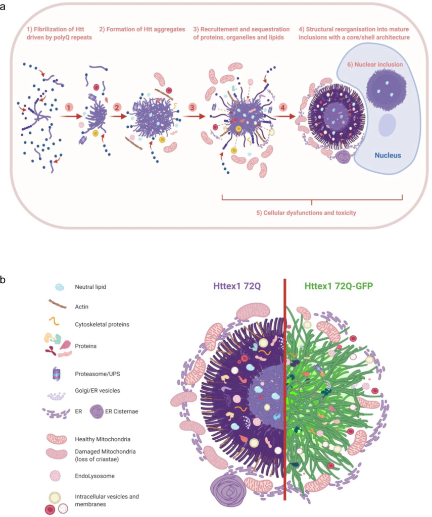
The formation of Huntingtin inclusions is driven primarily by the polyQ repeat domain and involves the active recruitment and sequestration of lipids, proteins, and membranous organelles.
a We separated the formation and organization of cytoplasmic Httex1 inclusions into different elements: (1) Even though the Nt17 domain can play a major role in early oligomerization steps, we showed that the fibrilization is driven by the polyQ repeats; (2) The initiation of inclusion formation was previously demonstrated to occur in a phase transition mechanism14,31 and could involve first the sequestration of proteins and organelles nearby either directly or indirectly; (3) During the growth of the inclusion, many cellular proteins, endomembranes, and lipids are recruited and sequestered inside the cytoplasmic inclusions; (4) Mature cytoplasmic Httex1 inclusions formed in cells display a core and shell structural organization if the polyQ length is relatively long (72Q) but not if it is close to the pathogenic threshold (39Q), even if inclusions are still formed in the second case. Cytoplasmic inclusion formation leads to the accumulation of mitochondria and ER network at the periphery; (5) Httex1 inclusion formation leads to the adaptation of the ER-mitochondrial network and respirational dysfunction, as well as significant toxicity after long-term incubation; (6) Nuclear inclusions do not display a distinct core/shell organization similar to the cytoplasmic inclusions, but they are still detected as a ring, as they are impervious for antibodies. Nuclear tag-free Httex1 inclusions are also enriched in neutral lipids. b The depiction shows the distinct structural organization and cellular impact of Httex1 72Q (left) compared to Httex1 72Q-GFP (right). The arrangement and packing of Httex1 fibrils are different depending on the presence of GFP as well as the interactions, recruitment, and perturbation with surrounding organelles.