
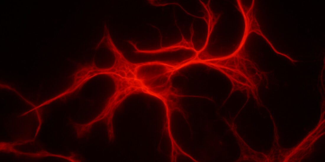
Astrocytes: the neglected stars in the central nervous system and drug addiction
By Wenjun Chen, Shiqiu Meng, Ying Han, and Jie Shi
Excerpt from the article published in Medical Review, vol. , no. , 2022. Published online: June 29, 2022 DOI: https://doi.org/10.1515/mr-2022-0006
Editor’s Highlights
- The star-shaped astrocyte, one dominant type of glia, has been implicated in myriad biological processes of the central nervous system (CNS) by interacting with almost all elements of the CNS, including neurons, synapses, glial cells and blood vessels.
- The astrocyte is widely distributed in the CNS. Its direct contact with neurons and blood vessels makes it a modulator in the neurovascular unit, forming the neuro-glial-vascular coupling, which is essential in regulating blood flow, substance and energy metabolism, and brain barrier construction.
- Drug addiction is a chronic relapsing disorder, that apart from endangering the health of individuals can also lead to crime and violence, and speed up the spread of infectious diseases, seriously affecting social stability and safety.
- There are some astrocyte-related molecules that participate in drug addiction, such as TSP2.
- By activating its neuronal receptor alpha2delta-1 (α2δ-1), astrocyte-secreted thrombospondins TSP2 could promote the generation of silent synapses.
- Cocaine triggers synaptogenesis in the nucleus accumbens (NAc) shell, and disrupting TSP2-α2δ-1 signaling could effectively prevent cue-induced relapse after extinction or withdrawal.
- A major obstacle in addiction treatment is the high rate of relapse due to the persistence of maladaptive drug-associated memories.
Abstract
With the advent of improved tools to examine the astrocytes, which have been believed to play a supportive role in the central nervous system (CNS) for years, their participation in the operation of the CNS and drug addiction was unveiled. Assisting the formation and function of the CNS, astrocytes are involved in physiological and pathological brain activities. Drug addiction is a pervasive psychiatric disorder, characterized by compulsive drug-taking behavior and high rate of relapse, impacting individual health and society stability and safety. When exposed to drugs of abuse, astrocytes go through a series of alterations, contributing to the development of addiction. Here we review how astrocytes contribute to the CNS and drug addiction. We hope that understanding the interaction between addictive drugs and astrocytes may help discover new mechanisms underlying the addiction and produce novel therapeutic treatments.
Introduction
The two mainstreams of cells that build up the central nervous system are neurons and glia, the latter of which for quite a long time are supposed to act as the background of the former, supporting, nourishing and protecting the neurons [1]. However actually, glia has much more functions. The star-shaped astrocyte, one dominant type of glia, has been implicated in myriad biological processes of the central nervous system (CNS) by interacting with almost all elements of the CNS, including neurons, synapses, glial cells and blood vessels [2]. Widely distributed in the whole brain, astrocytes function complexly varying from physiological activities to pathological changes.
Drug addiction is a chronic relapsing disorder, accounting for the loss of 18 million years of healthy life in 2019 [3]. According to World Drug Report 2021, it was estimated that about 36.3 million people were suffering from drug use disorder in 2019 and the number had rapidly accelerated year by year [3]. Apart from endangering the health of individuals, drug addiction can also lead to crime and violence, and speed up the spread of infectious diseases, seriously affecting social stability and safety [3]. Hence, it is of great significance to reveal the mechanism behind drug addiction, and thanks to the advancing technology in the neuroscience, accumulating studies help people understand how versatile astrocytes work in drug addiction indeed [4].
In the current review, we describe the astrocytic involvement in the operation of the CNS, and then focus on how astrocytes contribute to drug addiction through diversified pathways. Finally, we further discuss the existing problems and prospects of research in astrocytes and drug addiction.
How astrocytes contribute to the operation of the CNS
Astrocytes are involved in shaping the CNS
Neurogenesis and axon guidance
During the development of the CNS, astrocytes could secrete a variety of trophic factors to support neuronal life, such as brain-derived neurotrophic factor (BDNF), epidermal growth factor (EGF), fibroblast growth factor 2 (FGF‐2) and somatostatin [5–8]. In addition to promoting the growing process of neurons which are created earlier than glia, astrocytes mediate adult neurogenesis as well [9, 10]. Song et al. [11] first proved that astrocytes within adult hippocampus are able to accelerate the proliferation of stem cells and instruct them to become neurons. Following generation, neurons need to extend axons in order to establish synaptic connections, crossing long distances and complicated environment [12]. Astrocytes were also found to be critical for right pathfinding of axons. Minocha et al. [13] showed that Nkx2.1-positive astrocytes could guide axons through the expression of Slit2.
Synapse formation and synapse elimination
Since axons have reached the exact origin, neurons begin to form synapses with the help of astrocytes once again. In terms of the sequence of events, synaptogenesis happens right after the production of astrocytes and the time window of synapse formation overlaps that of the astrocyte maturation [14].
First of all, astrocytes make local contact with immature neurons, permitting them to receive and response to astrocyte-encoded signals. Switch of the receptivity may involve propagation of protein kinase C (PKC) signaling [15, 16]. The signals can be divided into prosynaptogenic ones and antisynaptogenic ones [17]. Prosynaptogenic signals include thrombospondins (TSP1 and TSP2) and Hevin, which could help construct the structure of synapses which contain presynaptic vesicles, N-methyl-D-aspartic acid receptors (NMDARs) and other elements [18, 19]. The synapses are presynaptically active; however, they are postsynaptically inactive so far, due to lack of α-amino-3-hydroxy-methyl-4-isoxazolepropionic acid receptors (AMPARs) [18]. Only with the aid of other prosynaptogenic signals, which could promote AMPARs localization to postsynaptic sites, such as glypicans 4 and 6, can synapses be fully functionally active [20]. Competitively, antisynaptogenic signals like secreted protein acidic rich in cysteine (SPARC) negatively regulate synapse formation [19]. Other astrocyte-secreted molecules also work during the synaptogenesis, including neuroligins, tumor necrosis factor-α (TNF-α), cholesterol, and transforming growth factor β-1 (TGFβ-1) [21–23].
Another important step in synaptic development is the elimination of weakened or redundant synapses, maintaining a proper number of synapses [23]. Astrocytes phagocytose excessive synapses in the developing brain through their phagocytic receptors multiple epidermal growth factor-like domains 10 (MEGF10) and Mer tyrosine kinase (MERTK) [24]. In the adult hippocampus, astrocytes continue to engulf impaired excitatory and inhibitory synapses for circuit homeostasis in an activity-dependent manner [25]. Hevin, which has been mentioned before in the synapse formation, is also a pivotal synapse refinement mediator. During early development, cortical dendritic spines often receive excitatory inputs from both cortex and thalamus, whereas Hevin can stabilize thalamic inputs, eventually forming single-input synapses [26]. On the contrary, in Hevin knockout mice, thalamic inputs are unable to compete with intracortical inputs, and thus ultimately resulting in a situation where the total number of cortical inputs increases and multiple excitatory input synapses persist [26]. Apart from direct mechanisms above, astrocytes can indirectly regulate synapse pruning through initiating microglia [27]. Astrocyte-secreted Interleukin-33 (IL-33) promotes microglial synapse engulfment during neural circuit maturation and remodeling [28].
All in all, astrocytes are involved in the formation of the CNS, assisting in the generation and development of neurons and regulating the synapse formation and elimination.
Astrocytes assist in the running of the CNS
Neuro-glial-vascular coupling
The astrocyte is widely distributed in the CNS. Its direct contact with neurons and blood vessels makes it a modulator in the neurovascular unit, forming the neuro-glial-vascular coupling, which is essential in regulating blood flow, substance and energy metabolism and brain barrier construction [29].
Reliable neural activity within the CNS demands strictly controlled environment and the blood-brain barrier (BBB) is a major interface that isolates brain compartment and circulating blood, adjusting the influx and efflux of solutes [30–32]. The astrocyte, with its endfeet physically ensheathing the capillaries, is located at a strategic position between neurons and endothelial cells [33]. Nutrients can be transported from blood to brain and waste compounds reversely via astrocytes [34, 35]. When the neuronal activity is enhanced, with abundant glutamate released, uptake of glucose by astrocytes from the bloodstream via glucose transporter type 1 (GLUT1) couples with the uptake of glutamate [36]. Through aerobic glycolysis, the glucose is converted to lactate which can be transferred by monocarboxylate transporters (MCT) from astrocytes to neurons [37]. Subsequently, lactate can be oxidized to pyruvate, which is utilized via the tricarboxylic acid cycle to produce vast adenosine triphosphate (ATP) to meet the great demand of energy.
Meanwhile, the endfeet which express water channel aquaporin 4 (AQP4) and Kir4.1 K+ channel play a special role in ion and volume regulation [31]. Potassium ions released from excited neurons reach astrocyte processes and then diffuse to the perivascular endfeet. Spreading K+ to a larger area achieves a spatial buffering effect [38]. Water influx, together with ion entry is tuned with water efflux through AQP4 [31]. The highly coordinated work of K+ channels and AQP4 realizes the clearance of extracellular K+ and balances ions and water in the microenvironment.
Homeostasis of the brain microenvironment
Aside from AQP4 and Kir4.1 at the endfeet, astrocytes have a mass of other transporters, channels, and enzymes to maintain the homeostasis of the brain microenvironment, such as ions, water, pH and neurotransmitters [39]. Besides the K+ channel, astrocytes can transfer K+ through transporters, such as Na+/K+ ATPase and Na+/K+/Cl− cotransporter 1, the former of which is also responsible for maintaining transmembrane Na+ gradient necessary for driving other transporters [40]. CO2 from neurons can be turned to HCO3 − by carbonic anhydrase and HCO3 − can be released by Na+/bicarbonate cotransporter to balance the pH [39]. Moreover, astrocytes remove and inactivate neurotransmitters, which include glutamate, norepinephrine, γ-aminobutyric acid (GABA) and adenosine, and release gliotransmitters like glutamate, ATP, D-Serine, as well as glutamine, an important source of glutamate and GABA [41].
Synaptic function and plasticity
There is a growing body of evidence that astrocytes are not merely supporting neurons, but also are intimately involved in the modulation of neuronal activity through bidirectional communication with synapses. Araque et al. [42] proposed a term ‘tripartite synapse’ to refer to the functional and physical structure of the presynaptic membrane, postsynaptic membrane and surrounding astrocyte. Unlike neurons, astrocytes show little electrical excitability, but their Ca2+ can be elevated as a result of activation [43]. In the ‘tripartite synapse’ model, elevation of astrocytic Ca2+level is triggered by neurotransmitters released during synaptic activation, and in turn, activated astrocytes release gliotransmitters to influence synaptic transmission [44]. It is estimated in rodent brain that a single astrocyte oversees 20 to 120 thousand synapses, making it in close relationship with synaptic function and plasticity [45].
Classic long-term potentiation (LTP) relies on NMDA receptors, whose activation needs binding of both glutamate and co-agonist D-serine, and astrocyte-derived D-serine modifies NMDAR plasticity in excitatory synapses nearby [46, 47]. Depletion of D-serine in an individual astrocyte blocks LTP formation, while supply of D-serine rescues LTP blockade induced by clamping astrocytic Ca2+ signals [47]. Astrocyte-derived glutamate occurring upon rise of Ca2+level could transiently increase the releasing probability of transmitter, and this short-term plasticity can be transformed to LTP due to the pairing of neuronal depolarization and astrocyte activation [48, 49]. ATP, which is rapidly converted to adenosine extracellularly, is another gliotransmitter. Astrocytes in the hippocampus CA1 region elicit ATP/adenosine, followed by upregulated basal synaptic transmission through presynaptic A2A receptors [50]. In the amygdala of mice, ATP/adenosine could even play different roles by depressing excitatory synapses via A1receptors and enhancing inhibitory synapses via A2A receptors [51]. Actually, a single astrocyte could release different gliotransmitters depending on the neuronal activity [52]. For instance, low frequency or short interneuron stimulation induces glutamate release from astrocytes leading to a short-term potentiation, whereas high frequency or prolonged stimulation also induces ATP/adenosine release leading to a short-term depression [52].
Additionally, morphology plasticity of astrocytes plays a key role in local synaptic activity through the structure perisynaptic astrocytic processes (PAPs). PAPs express a large amount of proteins relevant to synaptic transmission, including metabotropic glutamate receptors (mGluRs), glutamine synthetase, glutamate transporters, and GABABreceptors [45]. The extent to which astrocytes enwrap synapse elements affects the efficacy and activity of transmitter release, further influencing synaptic plasticity [2]. For example, in the supraoptic nucleus of lactating rats, the coverage of PAPs on synapses are reduced, leading to glutamate spillover and reduction of D-serine availability in the postsynaptic NMDARs and thus long-term synaptic changes [46]. A recent study shows that LTP induction prompts withdrawal of PAPs, which boosts extrasynaptic glutamate escape, therefore enhancing nearby synapses [53].
In summary, astrocytes mediate the function of the CNS, participating in neuro-glial-vascular coupling and maintaining the homeostasis of brain microenvironment. Besides, astrocytes are capable of detecting neuronal activity and playing an active role in modulating synaptic transmission.
How astrocytes contribute to drug addiction
Addictive drugs strongly activate dopamine signaling, and recent studies have also shown that astrocyte activity is fundamental for dopamine-evoked synapse regulation, suggesting that astrocytes may have an emerging role in drug addiction and may serve as a potential therapeutic target [54, 55].
GFAP expression and astrocyte morphology
Astrocytes can be identified by many markers, among which glial fibrillary acidic protein (GFAP) is upregulated after biological injury or during activation by harmful stimuli including drugs of abuse [56, 57]. Almost all kinds of addictive drugs could lead to changes in astrocyte GFAP expression and astrocyte morphology remodeling, yet it is difficult to define specific change patterns in distinct brain regions and in response to different drugs with various training paradigms [58–62].
After both acute and chronic cocaine exposure, GFAP elevation has been observed in the hippocampus [63]. Even after 7 days of cocaine treatment followed by a 3 week withdrawal period, GFAP is found to increase in the prefrontal cortex and both core and shell of the nucleus accumbens (NAc) [59]. However, in a model of cocaine self-administration and extinction training, Scofield et al. [64] pointed out reduction in GFAP expression in the NAc core, making it controversial on the effects of cocaine on changes in GFAP expression. In comparison, the majority of studies which have investigated GFAP expression following drug administration have reported increases consistently in amphetamine, methamphetamine and morphine [60–62]. Further, in human alcoholics, GFAP expression was significantly higher than controls in NAc [65]. Likewise, more GFAP is expressed in rat NAc core during abstinence from ethanol self-administration [66].
With regard to the morphology, astrocytes generally have a decrease in volume and length of processes after drug treatment. Chronic cocaine injection leads to reduced area and length of processes of dorsal hippocampal astrocytes [63]. In the NAc core, cocaine self-administration and extinction reduce astrocyte surface area and volume, as well as communication between astrocytes and synapses. In the same way, the extent of contact made by PAPs is decreased following methamphetamine self-administration and extinction [64, 67]. However, the reduction seems to be region-specific, for no differences in the prelimbic region of the medial prefrontal cortex and basolateral nucleus of the amygdala were observed [68]. As for nicotine, the condition goes totally different. In the prefrontal cortex, CA1 of the hippocampus and the substantia nigra, long-term exposure to nicotine induces extension of processes and increase of cell volume [69].
To sum up, drugs of abuse induce changes in astrocytes from GFAP expression to cell morphology, indicating that astrocytes perform certain functions in drug addiction. Although current outcomes demonstrate distinct responses to drugs, future studies are still needed to explore the influence of different kinds of drugs, administration doses and routes on the properties of astrocytes.
Maladaptive glutamatergic homeostasis
Among the maladaptive responses to addictive substances is the severely impaired glutamatergic homeostasis in the NAc, which is indispensable to the reinstatement of drug-seeking behavior induced by drug-associated cues, contexts, stress and drug itself [70–72].
Three crucial transporters or receptors are responsible for the astrocytic and synaptic glutamate release and elimination. They are glutamate transporter 1 (GLT-1), the cystine/glutamate exchanger (xCT) and mGluR2/3. After presynaptic membrane releases glutamate to postsynaptic membrane, astrocytes could remove extra glutamate from the synapse cleft through Na+-dependent GLT-1, which is in charge of more than 90% glutamate uptake in the brain [73]. Another transporter controlling extracellular glutamate levels is xCT, one of the several ways of astrocyte releasing glutamate, but the most influential one, through 1:1 exchange for extracellular cysteine, providing approximately 60% of the extrasynaptic glutamate in the NAc core [74]. Besides, mGluR2/3 distributed in presynaptic membranes could limit synaptic release of glutamate and its activation is related with xCT activity [75].
Upregulated glutamatergic transmission within the NAc underlies the reinstatement of drug-seeking behavior [59]. Numerous studies have shown that chronic exposure to drugs of abuse reduces the expression of GLT-1 and induces PAPs retraction, resulting in glutamate spillover in the cleft, accelerating that pathological process [75, 76]. In the same manner, the level of xCT is lowered after administration of drugs [77]. Accordingly, the reduced extracellular glutamate disinhibits regulation of presynaptic mGluR2/3 and enhances glutamate signaling, contributing to relapse behaviors [78]. Ceftriaxone or N-acetylcysteine that could restore expression of GLT-1 and xCT have proved to attenuate the reinstatement in cocaine, methamphetamine and heroin seeking [79–84]. Furthermore, chemogenetic activation of astrocytes by Gq-designer receptors exclusively activated by designer drugs (DREADDs) selectively drives astrocytes glutamate release and inhibits cue-induced cocaine seeking by stimulating mGluR2/3 [85]. Endocannabinoid signaling is another possible pathway to restore glutamate homeostasis, through which mGluR2/3 function is maintained and priming or cue-induced reinstatement of cocaine seeking is diminished [86].
To sum up, the disruption of glutamatergic homeostasis, which is strictly regulated by astrocytes, promotes vulnerability to reinstatement. Both the augmented synaptic glutamate release and reduced elimination from the synapse cleft could engender abnormal overflow of glutamate linked to the reinstatement of drug seeking behavior.
Astrocyte-neuron signaling in drug addiction
Under physiological circumstances, a huge number of gliotransmitters, transporters and receptors of astrocytes are conducive to its regulation to neuronal activity. Under the pathological conditions of drug abuse, these molecules also work in the reciprocal crosstalk between astrocytes and neurons.
Addictive drugs, despite of distinct action mechanisms, activate the dopamine system consistently [87]. Recently, Corkrum et al. [55] showed that astrocytes in the NAc mediate dopamine-induced synaptic depression through D1 receptors. With elevation of intracellular Ca2+ level, activated astrocytes release ATP/adenosine binding to presynaptic A1 receptors to depress excitatory synapse transmission, which is necessary for amphetamine-related manifestations [55]. Astrocytic adenosine signaling has a significant role in the transition from habitual to goal-directed reward-seeking behavior and alcohol-seeking behavior [88, 89]. In the early stage of drug abuse, drug seeking is controlled and goal-directed, but it gradually turns habitual and compulsive as the addiction develops [90]. Kang et al. [88] demonstrated that chemogenetic activation of astrocytes in the dorsomedial striatum regulates the activity of medium spiny neurons, shifting the reward-seeking behavior patterns from habitual actions to goal-directed ones via adenosine signaling.
Astrocyte-derived D-serine, closely related to synapse plasticity, is also a signal molecule of drug addiction. Curcio et al. [91] proved that exposure to cocaine results in reduced D-serine levels, and therefore subsequently impaired NMDAR-dependent potentiation and depression of glutamatergic synaptic transmission in the NAc. For this reason, exogenous D-serine supply succeeds in rescuing the damaged plasticity and interfering cocaine-related behaviors. As revealed by Kelamangalath & Wagner [92], D-serine treatment facilitates the extinction training and attenuates cocaine-primed drug-seeking behavior. Same effects have been found in cocaine-induced conditioned place preference (CPP) and locomotor sensitization [93, 94]. Moreover, morphine inhibits D-serine release from astrocytes, suppressing the excitability of postsynaptic GABAergic neurons [95].
Lactate is a novel mediator in learning and memory, synapse plasticity and drug addiction, which has been considered only as an energy substrate in brain energy metabolism for long [36]. Since Suzuki et al. [96] found astrocyte-neuron lactate transport is of great importance for long-term memory formation, its role in drug memory, which is associated with drug abuse and relapse, has aroused lots of attention of scientists. Pharmacological inhibition of glycogenolysis in the basolateral amygdala disrupts the lactate production, along with the impaired acquisition and persistence of cocaine-induced CPP [97]. Meanwhile, disruption of astrocyte-neuron lactate transport abolishes the reconsolidation of cocaine reward memory and subsequent expression of cocaine-induced CPP [98]. Lactate is also supposed to be involved in glucocorticoid receptor (GR) –mediated alterations in synapse transmission caused by morphine [99]. While astrocytic GR knockdown inhibits glucocorticoid-induced lactate release, it enhances morphine-induced CPP at the same time, and lactate supplementation could reverse that action [99].
There are still some other astrocyte-related molecules that participate in drug addiction, such as TSP2 and AQP4. By activating its neuronal receptor α2δ-1, astrocyte-secreted TSP2 could promote generation of silent synapses [18]. Through this way, cocaine triggers synaptogenesis in the NAc shell, and disrupting TSP2-α2δ-1 signaling could effectively prevent cue-induced relapse after extinction or withdrawal [100]. Furthermore, ablation of AQP4 reduces heroin consumption in self-administration training and morphine-induced behavioral sensitization, which may be achieved by upregulated expression of dopamine transporter [101].
In conclusion, astrocytes could affect neuronal and synaptic activity through multiple ways to respond to the development of drug addiction. There’s no doubt that astrocyte-neuron interaction via glutamate, ATP/adenosine, D-serine, lactate etc., is an essential mechanism of drug addiction.
Future perspectives
For decades, the main character in the neuroscience research has always been the neuron, whose electrical activity is considered to be the foundation of brain activity, while glia plays a secondary and supportive role. However, improved techniques to visualize and manipulate glia have thoroughly expanded our knowledge of glial functions in physiological and pathological conditions, especially those of the astrocytes, the most abundant glial cells [102]. Fruitful findings have prompted us to appreciate the prominent role of astrocytes in the CNS. In terms of shaping the CNS, astrocytes assist neurogenesis and axon guidance, as well as controlling an appropriate number of synapses by participating in synapse formation and elimination. As for the CNS function, astrocytes own diversified receptors, transporters, channels, enzymes and gliotransmitters involved in neuro-glial-vascular coupling, microenvironment homeostasis maintenance, synapse plasticity regulation and many others. These gratifying progresses allow us to re-examine the nervous system and neuropsychiatric diseases from a different perspective.
Since the past few years have witnessed inspiring achievements in acknowledging the function of astrocytes, people began to explore their roles in drug addiction. Most drugs of abuse could activate the astrocytes and alter their morphology and functions towards aberrant levels, which contributes to the development of maladaptive drug-related behaviors. Specifically, the patterns of astrocyte releasing gliotransmitters, such as glutamate, ATP/adenosine and D-serine, are impacted. Among those substances, glutamate has been studied extensively in particular, for the disruption of its homeostasis is believed to promote vulnerability to relapse.
Better understanding of the molecular and cellular changes induced by drugs allows the seeking of effective treatments for drug addiction, among which manipulating astrocytes has risen as a unique approach to prevent relapse. Although growing evidence has demonstrated that some pharmacological means of restoring glutamatergic homeostasis can successfully prevent relapse in rodents, the results of clinical trials in humans are still limited. N-acetylcysteine treatment could potently reduce drug desire following either cocaine cue exhibition or intravenous cocaine injection [103, 104]. In another trial, N-acetylcysteine administration failed to reduce cocaine use in active users, but was able to decrease drug craving in those who had achieved abstinence [105]. Combined with cognitive behavioral therapy, N-acetylcysteine treatment for 8 weeks significantly lessened the craving in veterans with substance use disorder [106]. Recent findings substantiate that N-acetylcysteine could attenuate cocaine-cue attentional bias by reducing the incentive salience of cocaine, and decrease cocaine-seeking behavior possibly by modulating glutamate levels in the rostral anterior cingulate [107, 108]. Although above results are inconsistent, N-acetylcysteine, which is well-tolerated with only mildly adverse effects, displays its potential in preventing drug relapse [109]. Besides N-acetylcysteine, in methamphetamine users, electroacupuncture has the ability to normalize glutamate levels by enhancing astrocytic glutamate clearance in the dorsal hippocampal CA1, suggesting that non-invasive electroacupuncture might be a novel approach to manage drug addiction [110].
As a matter of fact, drug addiction is also a disorder of learning and memory [111]. A major obstacle in addiction treatment is the high rate of relapse due to the persistence of maladaptive drug-associated memories [112]. Even after a long period of withdrawal, individuals have a propensity to generate drug-taking and drug-seeking behaviors when exposed to drug-associated cues and environment [113]. Recently, converging evidence of astrocytic role in memory has been obtained with advanced techniques (including translating ribosome affinity purification, RiboTag, single-cell transcriptomic analyses, super-resolution microscopy, neuron-astrocyte proximity assay, etc. [4]), indicating that astrocyte may also play a part in drug memory as well, which may open a completely new area in pathological mechanism and clinical treatment research [25, 114–116].
In short, it has lately come to light that astrocytes are key participants in drug addiction and they are emerging as a promising therapeutic target of drug addiction.
Conclusions
Here, we highlighted diverse roles of astrocytes in many aspects of the CNS and drug addiction. Notwithstanding that extensive efforts have been made to disentangle the interaction between astrocytes and addictive drugs, further precise exploration in the circuit, cellular, molecular and genetic mechanism of astrocyte-mediated addiction is still needed. With the emergence of new strategies to interrogate astrocytes in vivo, a more comprehensive understanding of the roles of astrocytes in drug addiction will be achieved, holding considerable promise for developing feasible therapeutic treatments of drug addiction in the future.