
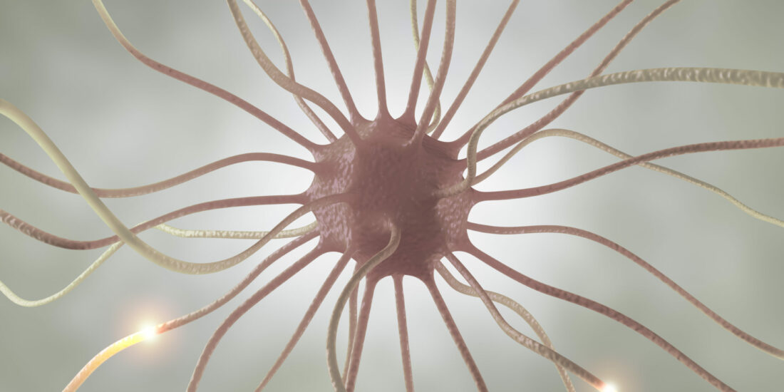
Presynaptic α2δ subunits are key organizers of glutamatergic synapses
By Clemens L. Schöpf, Cornelia Ablinger, Stefanie M. Geisler, Ruslan I. Stanika, Marta Campiglio, Walter A. Kaufmann, Benedikt Nimmervoll, Bettina Schlick, Johannes Brockhaus, Markus Missler, Ryuichi Shigemoto, and Gerald J. Obermair
Excerpt from the article published in PNAS (Proceedings of the National Academy of Sciences) March 29, 2021 | 118 (14) e1920827118 | https://doi.org/10.1073/pnas.1920827118
Editor’s Highlights
- The extracellular structure of α2δ-1 of the skeletal muscle CaV1.1 complex proposes the protrusion of α2δ subunits far into the synaptic cleft.
- Postsynaptic α2δ-1 acts as a receptor for thrombospondins and promotes spinogenesis via postsynaptic Rac1.
- The antiepileptic and antiallodynic drug gabapentin prevents synaptogenesis between sensory and spinal cord neurons by acting on presynaptic α2δ-1 subunits.
Significance
Voltage-gated calcium channels are important regulators of neuronal functions, as for example synaptic transmission. Their auxiliary α2δ subunits are modulating the calcium currents. Beyond that they have emerged as modulators of synaptic functions. Here, we established a cellular triple knockout/knockdown model in cultured hippocampal neurons by knocking out or knocking down the expression of all three α2δ subunits expressed in brain. Our experiments demonstrate that the presynaptic loss of α2δ proteins leads to a severe defect in glutamatergic synapse formation, which could be rescued by reintroducing any of the three neuronal α2δ isoforms. Thus, our study suggests that α2δ proteins are critical regulators of excitatory synapse formation and thereby contributes to the understanding of basic nerve cell functions.
Abstract
In nerve cells the genes encoding for α2δ subunits of voltage-gated calcium channels have been linked to synaptic functions and neurological disease. Here we show that α2δ subunits are essential for the formation and organization of glutamatergic synapses. Using a cellular α2δ subunit triple-knockout/knockdown model, we demonstrate a failure in presynaptic differentiation evidenced by defective presynaptic calcium channel clustering and calcium influx, smaller presynaptic active zones, and a strongly reduced accumulation of presynaptic vesicle-associated proteins (synapsin and vGLUT). The presynaptic defect is associated with the downscaling of postsynaptic AMPA receptors and the postsynaptic density. The role of α2δ isoforms as synaptic organizers is highly redundant, as each individual α2δ isoform can rescue presynaptic calcium channel trafficking and expression of synaptic proteins. Moreover, α2δ-2 and α2δ-3 with mutated metal ion-dependent adhesion sites can fully rescue presynaptic synapsin expression but only partially calcium channel trafficking, suggesting that the regulatory role of α2δ subunits is independent from its role as a calcium channel subunit. Our findings influence the current view on excitatory synapse formation. First, our study suggests that postsynaptic differentiation is secondary to presynaptic differentiation. Second, the dependence of presynaptic differentiation on α2δ implicates α2δ subunits as potential nucleation points for the organization of synapses. Finally, our results suggest that α2δ subunits act as transsynaptic organizers of glutamatergic synapses, thereby aligning the synaptic active zone with the postsynaptic density.
In synapses neurotransmitter release is triggered by the entry of calcium through voltage-gated calcium channels (VGCCs). Neuronal VGCCs consist of an ion-conducting α1 subunit and the auxiliary β and α2δ subunits. α2δ subunits, the targets of the widely prescribed antiepileptic and antiallodynic drugs gabapentin and pregabalin, are membrane-anchored extracellular glycoproteins, which modulate VGCC trafficking and calcium currents (1–5). In nerve cells α2δ subunits have been linked to neuropathic pain and epilepsy (4) and they interact with mutant prion proteins (6) and regulate synaptic release probability (7). Importantly, all α2δ isoforms are implicated in synaptic functions. Presynaptic effects of α2δ-1, for example, may be mediated by an interaction with α-neurexins (8) or N-methyl-D-aspartate receptors (e.g., refs. 9 and 10). In contrast, postsynaptic α2δ-1 acts as a receptor for thrombospondins (11) and promotes spinogenesis via postsynaptic Rac1 (12). α2δ-2 is necessary for normal structure and function of auditory hair cell synapses (13); it has been identified as a regulator of axon growth and hence a suppressor of axonal regeneration (14) and was recently shown to control structure and function of cerebellar climbing fiber synapses (15). A splice variant of α2δ-2 regulates postsynaptic GABAA receptor (GABAAR) abundance and axonal wiring (16). In invertebrates, α2δ loss of function was associated with abnormal presynaptic development in motoneurons (17, 18) and in mice the loss of α2δ-3 results in aberrant synapse formation of auditory nerve fibers (19). Finally, α2δ-4 is required for the organization of rod and cone photoreceptor synapses (20, 21).Despite these important functions, knockout mice for α2δ-1 and α2δ-3 show only mild neurological phenotypes (5, 10, 22–25). In contrast, mutant mice for α2δ-2 (ducky) display impaired gait, ataxia, and epileptic seizures (26), all phenotypes consistent with a cerebellar dysfunction due to the predominant expression of α2δ-2 in the cerebellum (e.g., ref. 15). Hence, in contrast to the specific functions of α2δ isoforms (discussed above) the phenotypes of the available knockout or mutant mouse models suggest a partial functional redundancy in central neurons. Moreover, detailed mechanistic insights into the putative synaptic functions of α2δ subunits are complicated by the simultaneous and strong expression of three isoforms (α2δ-1 to -3) in neurons of the central nervous system (27).In this study, by transfecting cultured hippocampal neurons from α2δ-2/-3 double-knockout mice with short hairpin RNA (shRNA) against α2δ-1, we developed a cellular α2δ subunit triple-knockout/knockdown model. Excitatory synapses from these cultures show a severe failure of synaptic vesicle recycling associated with severely reduced presynaptic calcium transients, loss of presynaptic calcium channels and presynaptic vesicle-associated proteins, and a reduced size of the presynaptic active zone (AZ). Lack of presynaptic α2δ subunits also induces a failure of postsynaptic PSD-95 and AMPA receptor (AMPAR) localization and a thinning of the postsynaptic density (PSD). Each individual α2δ isoform (α2δ-1 to -3) could rescue the severe phenotype, revealing the highly redundant role of presynaptic α2δ isoforms in glutamatergic synapse formation and differentiation. Together our results show that α2δ subunits regulate presynaptic differentiation as well as the transsynaptic alignment of postsynaptic receptors and are thus critical for the function of glutamatergic synapses.
Results
Epitope-Tagged α2δ Isoforms Localize to Presynaptic Boutons.
Three isoforms of the calcium channel α2δ subunit are expressed in hippocampal neurons (27), yet until today it is unclear whether all three isoforms contribute to specific neuronal and synaptic functions. A differential subcellular compartmentalization of α2δ isoforms could provide insights into their specific functions. Therefore, we first investigated the localization of hemagglutinin (HA)-epitope-tagged α2δ-1, -2, and -3 in cultured hippocampal neurons. To this end a double HA-tag was engineered into N termini of all three α2δ subunits cloned from mouse brain complementary DNA (GenBank accession numbers MK327276, MK327277, and MK327280) right after the signal sequence. Live-cell immunolabeling of the HA-epitope allows a direct and, most importantly, comparative analysis of α2δ isoform surface expression. Using the same antibody (anti-HA) for quantitatively comparing distinct α2δ isoforms provides an important advantage over currently available α2δ antibodies, which either do not reliably detect the native proteins (28) or are not suitable for immunocytochemical experiments (29). Although the overall intensity of total surface expression levels differs between isoforms (α2δ-2 > α2δ-3 > α2δ-1), all three isoforms are localized to the somatodendritic and axonal membrane (SI Appendix, Fig. S1A). In addition, α2δ-3 shows a preferential expression in the axon. However, despite these apparent overall differences all α2δ isoforms are expressed on the surface of axons and presynaptic membranes (SI Appendix, Fig. S1B), suggesting that, in principle, all three isoforms can contribute to synaptic functions.
α2δ Subunit Isoforms Are Essential for Survival.
With the exception of the α2δ-2 mutant mouse ducky, knockout mice for α2δ-1 and α2δ-3 display only mild neuronal phenotypes, suggesting a potential and at least partial functional redundancy (discussed above). Therefore, in order to gain insight into the functional diversity of α2δ subunits, we generated double-knockout mice by pairwise cross-breeding single-knockout (α2δ-1 and α2δ-3) and mutant (α2δ-2du) mice (29). While α2δ-1/-3 knockout mice are viable for up to 3 mo, similar to ducky mice, α2δ-1/-2 and α2δ-2/-3 knockout mice have a strongly reduced lifespan (Fig. 1 A and B). A significant proportion of these mice require application of humane endpoints within the first postnatal week, mainly due to malnutrition associated with a poor general condition. Together this shows that α2δ subunits serve essential functions and are necessary for survival. Moreover, the increased severity of the phenotype in double- compared with single-knockout mice also supports the idea that α2δ subunits act in part redundantly.

Establishing a Cellular α2δ-Subunit Triple-Knockout/Knockdown Model.
In order to study a potential functional redundancy of α2δ subunits we next developed a cellular α2δ triple-knockout/knockdown model system by transfecting cultured hippocampal neurons from α2δ-2/-3 double-knockout mice with shRNA against α2δ-1. To this end we first confirmed efficient shRNA knockdown of α2δ-1 in two independent experimental settings. First, shRNA against α2δ-1 (30, 31) significantly reduced the surface expression of a heterologously expressed α2δ-1 isoform bearing an extracellular phluorin-tag (superecliptic phluorin, SEP; SI Appendix, Fig. S2 A and B); however, due to the experimental overexpression low levels of this α2δ-1 isoform were still detectable (37% of control; SI Appendix, Fig. S2 A and B). Second, qPCR analysis of cultured hippocampal neurons virally infected with α2δ-1 shRNA revealed an overall 80% knockdown of α2δ-1 messenger RNA (mRNA) compared with untransfected (wild-type) neurons or neurons expressing scrambled control shRNA (SI Appendix, Fig. S2C). Considering a ∼90% infection efficiency, confirmed by enhanced green fluorescent protein (eGFP) expression from the same viral vector, shRNA robustly knocked down mRNA in the vast majority of infected neurons. Most importantly, shRNA knockdown of α2δ-1 did not affect the expression levels of the other α2δ isoforms (SI Appendix, Fig. S2C). In order to evaluate potential compensatory mechanisms, we also quantified mRNA levels of all α2δ isoforms in hippocampal tissue from 8-wk-old single-knockout mice. Similar to α2δ-1 knockdown, neither loss of α2δ-2 nor of α2δ-3 induced compensational changes in the expression levels of the other isoforms (SI Appendix, Fig. S2 D and E).α2δ-2/-3 double-knockout mice were generated by cross-breeding double heterozygous α2δ-2+/du/α2δ-3+/− mice (SI Appendix, Fig. S3A). The predicted Mendelian ratio for double-knockout mice is 6.25%, however, the experimentally determined ratio was only ∼3% (see legend of SI Appendix, Fig. S3). Neonatal pups (postnatal day [P] 0 to 2) were individually marked by paw tattooing and genotyped for the α2δ-2 and α2δ-3 alleles (SI Appendix, Fig. S3 Band C). Due to the large genomic rearrangement in ducky mice, genotyping of the ducky mutation required a confirmation employing a copy-number-counting qPCR approach (SI Appendix, Fig. S3D). Ultimately, α2δ triple loss-of-function hippocampal neurons were established by transfecting confirmed α2δ-2/-3 double-knockout cultures with α2δ-1 shRNA and eGFP (SI Appendix, Fig. S3E).
Failure of Presynaptic Differentiation in α2δ Subunit Triple-Knockout/Knockdown Neurons.
In cultured hippocampal neurons from α2δ-2/-3 double-knockout mice, shRNA-transfected neurons (α2δ-2/-3 double-knockout with α2δ-1 shRNA knockdown, further referred to as α2δ TKO/KD) can be easily identified by the expression of soluble eGFP. Most importantly, in this experimental setting isolated axons and synaptic varicosities from transfected α2δ TKO/KD neurons can be directly compared with untransfected neighboring neurons, which still express α2δ-1 (SI Appendix, Fig. S4). Axons from cultured hippocampal neurons display axonal varicosities which can be morphologically identified by the eGFP fluorescence. Such varicosities are typically representing presynaptic boutons, as confirmed by the clustering of presynaptic proteins (e.g., CaV2.1 channels and synapsin; SI Appendix, Fig. S4, Left). Axons from α2δ TKO/KD neurons display similar axonal varicosities, mostly found located along dendritic processes of nontransfected neighboring double-knockout cells (SI Appendix, Fig. S4, Right). In order to test whether these boutons represent functional synapses capable of vesicle recycling we quantified the extent of depolarization-induced uptake of the styryl membrane dye FM4-64. Upon a 60 mM [K+]-induced depolarization 68% of the axonal varicosities of α2δ TKO/KD neurons completely failed to take up FM dye and loading of the remaining 32% was strongly decreased (Fig. 1C). In contrast, neighboring untransfected (α2δ-1–containing) synapses (Fig. 1 C, Right, asterisks) and eGFP-transfected control neurons were readily stained with FM4-64 upon high [K+] treatment. This apparent failure of synaptic vesicle recycling pointed toward a severe defect in presynaptic calcium channel functions.Indeed, voltage-clamp analysis of total somatic calcium currents identified a marked reduction of current densities by 58% (Fig. 1 D and E) and of the maximal conductance by 37% (SI Appendix, Fig. S5) but no change in the voltage-dependence of activation and half-maximal activation of α2δ TKO/KD compared with α2δ-3 single-knockout or wild-type control neurons (Fig. 1 D and E and SI Appendix, Table S1 and Fig. S5). Notably, current densities and maximal conductance were also reduced in α2δ-2/-3 double-knockout neurons (by 32% and 23%, respectively; Fig. 1E and SI Appendix, Fig. S5C), however without a concomitant failure in FM dye uptake (Fig. 1C). The homologous reconstitution of α2δ-2 in TKO/KD neurons fully rescued the currents back to wild-type levels (Fig. 1 D and E and SI Appendix, Table S1), while the sole presence of α2δ-1 in the α2δ-2/-3 double-knockout condition could not fully compensate the effects on total somatodendritic currents (Fig. 1D, red trace). Importantly, to avoid any possible influence of the HA-epitope tag (discussed above) on α2δ isoform function, all rescue experiments were performed with wild-type, untagged α2δ subunits.While reduced somatic calcium channel activity was to be expected in an α2δ-null model, the complete failure of FM dye uptake suggests a more severe failure of synaptic vesicle recycling. Therefore, we next tested the consequences of α2δ TKO/KD on presynaptic calcium signals. To this end neurons were transfected with the genetically encoded calcium indicator GCaMP6f, coupled to synaptophysin and expressed under the control of a synapsin promoter (32), together with soluble mCherry to outline neuronal and axonal morphology (Fig. 2A). Presynaptic calcium signals (ΔF/F0) were recorded and analyzed in putative presynaptic boutons (axonal varicosities, discussed above) in response to field stimulation triggering 1, 3, or 10 action potentials (APs) at a frequency of 50 Hz (Fig. 2B and Movies S1 and S2). α2δ TKO/KD resulted in a reduction of mean peak amplitudes to 15%, 34%, and 48% in response to 1, 3, and 10 APs, respectively, in comparison with double-heterozygous control neurons (Fig. 2 B and D). Ectopic overexpression of α2δ-1, which was reduced but not eliminated by shRNA (SI Appendix, Fig. S2B), rescued mean peak amplitudes to 117%, 104%, and 107% of control. Even more strikingly, frequency distribution histograms of peak responses of all synapses (Fig. 2C) show that 73%, 44%, and 39% of TKO/KD boutons failed to show any calcium transient in response to 1, 3, and 10 APs. In contrast, failures of heterozygous control neurons were observed in only 24%, 8%, and 5%, and those of α2δ-1-rescued neurons in only 43%, 23%, and 12% of synapses, each in response to 1, 3, and 10 APs, respectively. Together these results demonstrate a severe reduction of presynaptic calcium influx and hence suggest a defect in presynaptic calcium channel clustering in α2δ TKO/KD boutons.

Therefore, we next employed immunocytochemistry to test whether and to what extent the synaptic localization of presynaptic P/Q- (CaV2.1) and N-type (CaV2.2) calcium channels was affected (Fig. 3 A and B). Strikingly, 61% and 40% of the axonal TKO/KD varicosities lacked detectable staining for CaV2.1 and CaV2.2, respectively. The remaining axonal boutons showed a strong and significant reduction of presynaptic labeling intensities (Fig. 3 C and D). In agreement with defective synaptic vesicle recycling, these boutons were also deficient in synapsin staining (complete loss in 45% of the analyzed boutons; Fig. 3E). The strongly reduced presynaptic calcium channel abundance in α2δ TKO/KD varicosities is in line with the major role of α2δ subunits in enhancing calcium channel trafficking (33). However, the surprising loss of synapsin staining suggests that the lack of α2δ subunits also grossly affects presynaptic differentiation.

Presynaptic α2δ Subunits Regulate Pre- and Postsynaptic Differentiation of Excitatory Glutamatergic Synapses.
By acting as a thrombospondin receptor, α2δ-1 has previously been suggested to contribute to synaptogenesis by a postsynaptic mechanism (11, 12). Therefore, in order to distinguish between the proposed postsynaptic mechanism and the defect in presynaptic differentiation observed here, we examined α2δ TKO/KD neurons connected to neighboring nontransfected double-knockout neurons still expressing α2δ-1 (SI Appendix, Fig. S4). In this experimental paradigm, eGFP-positive axonal processes of presynaptic TKO/KD neurons (Fig. 4 A and B, Leftand sketches) can be clearly distinguished from eGFP-positive dendrites of postsynaptic TKO/KD neurons (Fig. 4 A and B, Right and sketches). These experiments demonstrate that synapse differentiation fails when the presynaptic neuron lacks all α2δ subunits (Fig. 4 A and B, Left). On the other hand, postsynaptic α2δ TKO/KD neurons can still form dendritic spines and receive synaptic inputs from neighboring double-knockout neurons expressing α2δ-1 (Fig. 4 Aand B, Right). This observation is confirmed by recording miniature excitatory postsynaptic currents (mEPSC) from postsynaptic α2δ TKO/KD neurons in comparison with heterozygous control or double-knockout neurons (Fig. 4 C and D). Neither mEPSC frequency nor amplitude is reduced in putative TKO/KD synapses, strengthening the conclusion that postsynaptic α2δ TKO/KD neurons can still receive synaptic inputs from neighboring double-knockout neurons.

he presynaptic defect in synapse formation also induced a failure in the postsynaptic differentiation: Boutons devoid of calcium channels or synapsin were either not juxtaposed to PSD-95 clusters at all (Fig. 4E) or the PSD-95 labeling was strongly reduced (Fig. 4F). Similar to the marked reduction of presynaptic synapsin and calcium channel labeling, PSD-95 was completely absent in 58% of the analyzed α2δ TKO/KD synapses. Thus, in addition to the failure in presynaptic differentiation, the lack of presynaptic α2δ subunits also induced a failure in postsynaptic differentiation. For analyzing whether presynaptic α2δ subunits are required for both excitatory and inhibitory synapse formation, we immunolabeled α2δ TKO/KD and control neurons for respective components of the presynaptic vesicle compartment and postsynaptic receptors (Fig. 4 G and H). In excitatory glutamatergic neurons the lack of presynaptic staining for the vesicular glutamate transporter type 1 (vGLUT1; for quantification see SI Appendix, Fig. S6) goes along with strongly reduced clustering of postsynaptic AMPARs in TKO/KD synapses (Fig. 4 G and I). Conversely, α2δ TKO/KD synapses from GABAergic neurons still seem to express the presynaptic vesicular GABA transporter (vGAT) and display postsynaptic clustering of GABAAR (Fig. 4 H and J). However, it is important to note that due to the low abundance of GABAergic neurons (∼5 to 10% of all cultured hippocampal neurons), the extremely low availability of α2δ-2/-3 double-knockout offspring (on average only two to five culture preparations are possible per year), and the necessity of shRNA transfection, we could only analyze two cells for control and five cells for TKO/KD conditions. Therefore, to confirm this finding we also analyzed the abundance of pre- and postsynaptic proteins in control (α2δ-3 knockout) and α2δ TKO/KD cultured GABAergic striatal medium spiny neurons (Fig. 5) (16). In contrast to glutamatergic hippocampal neurons, α2δ TKO/KD in GABAergic medium spiny neurons does not affect the abundance of presynaptic vGAT (Fig. 5D) and synapsin (Fig. 5H), the presynaptic bouton size (SI Appendix, Fig. S7), or postsynaptic GABAA-receptor clustering (Fig. 5C and SI Appendix, Fig. S7). However, although not statistically significant, there was a tendency for reduced presynaptic CaV2.1 labeling (Fig. 5G). Together this demonstrates that the severe consequence of presynaptic α2δ TKO/KD is specific to excitatory glutamatergic neurons.

α2δ Subunit Triple Knockout/Knockdown Affects the Pre- and Postsynaptic Ultrastructure.
Immunofluorescence labeling identified a strong reduction in the abundance of presynaptic and postsynaptic proteins in glutamatergic synapses of α2δ TKO/KD neurons. In order to test whether these presynaptic effects are associated with ultrastructural alterations we performed classical transmission electron microscopy (TEM) and preembedding immunoelectron microscopy. Classical TEM analysis revealed the necessity for immunolabeling shRNA-α2δ-1/eGFP transfected double-knockout neurons in order to reliably identify the sparsely distributed α2δ TKO/KD synapses weak in morphological cues. The strong immunolabeling for eGFP with the contrast intense silver-amplified gold particles, however, obscured the presynaptic ultrastructure and rendered reliable analysis of synaptic vesicle content and localization impossible. For quantifying size and extension of synaptic specializations, we first compared synapses of nonlabeled wild-type control and α2δ-2/-3 double-knockout neurons (Fig. 6A). Analyses of 40 synapses in each condition revealed that the length of the AZ and the PSD, the AZ/PSD ratio, as well as the PSD thickness (extension from the membrane into the cytosol) were indistinguishable between control and double-knockout neurons (mean ± SEM in nanometers, unpaired t test; AZ: control, 433 ± 22, double-KO, 433 ± 20, P = 0.99; PSD: control, 440 ± 23, double-KO, 436 ± 20, P = 0.89; PSD extension: control, 28.5 ± 1.4; double-KO, 27.2 ± 1.1, P = 0.49; AZ/PSD ratio: control, 0.986 ± 0.004, double-KO, 0.995 ± 0.006; P = 0.19). We next performed the same analysis on eGFP-immunostained double-knockout (control eGFP) and TKO/KD (triple KO) synapses (Fig. 6B). As an additional control, we measured the respective AZ and PSD parameters of nontransfected neighboring synapses (control nt), which are all double-knockout for α2δ-2/-3. Both AZ and PSD lengths were significantly reduced by ∼25% in α2δ TKO/KD synapses (Fig. 6 C, Left and Middle); however, the AZ/PSD ratio was not altered [AZ/PSD ratio: control eGFP, 1.00 ± 0.01, triple KO, 1.05 ± 0.04; control nt, 0.98 ± 0.04; ANOVA, F(2,147) = 1.45, P = 0.24]. This suggests that reductions in the presynaptic AZ caused by lack of α2δ subunits are directly affecting the size of the PSD. Control measurements in nontransfected synapses (control nt) were indistinguishable from eGFP-transfected α2δ-2/-3 double-knockout neurons (control eGFP). The extension of the PSD from the synaptic membrane into the cytosol was reduced by 40% in TKO/KD synapses compared with both controls (Fig. 6 C, Right). Taken together, these measurements reveal that presynaptic α2δ TKO/KD reduces the sizes of the presynaptic AZ and PSD as well as the thickness of the PSD (Fig. 4E).

The α2δ Subunit Triple-Knockout/Knockdown Phenotype Can Be Rescued by Overexpression of α2δ-1, -2, and -3.
The severe consequences of presynaptic α2δ triple loss of function on pre- and postsynaptic composition and synaptic ultrastructure strongly suggest a functional redundancy. Thus, to further elucidate the potentially redundant roles of α2δ subunits in pre- and postsynaptic differentiation, we analyzed the propensity of each individual isoform in rescuing synapse formation and differentiation. First, α2δ-2/-3 double-knockout neurons which solely express α2δ-1 showed a proper apposition of pre- and postsynaptic proteins (see colocalized synaptic markers near the eGFP-positive TKO/KD axons indicated by asterisks in Figs. 1C, 3 A and B, and 4 B and E). Moreover, the α2δ TKO/KD phenotype could be rescued by the expression of α2δ-2 (rescue in Figs. 1 D and E, 3, 4E, and 7 and SI Appendix, Fig. S5), α2δ-1 (SI Appendix, Figs. S6 and S8), and α2δ-3 (Fig. 7 and SI Appendix, Fig. S8). Together this shows that the apparent critical roles of α2δ subunits in glutamatergic synapse formation are highly redundant between the neuronal α2δ isoforms.

Expressing α2δ-ΔMIDAS Mutants in Triple-Knockout/Knockdown Neurons Fully Rescues Presynaptic Synapsin but Not Calcium Channel Clustering.
Our experiments demonstrate an essential role of α2δ subunits in glutamatergic synapse formation and differentiation which might be related to the failure of presynaptic calcium channel trafficking. Alternatively, however, α2δ subunits may act transsynaptically and independent of the calcium channel complex, as has been previously suggested (5, 13, 16, 18). α2δ subunits contain a von Willebrand factor type A (VWA) domain which, at least in α2δ-1 and α2δ-2, includes a perfect metal ion-dependent adhesion site (MIDAS). The integrity of the MIDAS motif in α2δ subunits is necessary for calcium current enhancement and channel trafficking (7, 34, 35). This finding is supported by the proposed structure of α2δ-1, in which the VWA domain and particularly the MIDAS are facing the surface of the pore-forming α1 subunit and are thus predicted to be crucial for α1 and α2δ subunit interactions (36). We reasoned that mutating the MIDAS site, which has previously been shown to inhibit channel trafficking (35), may be helpful in dissociating channel-dependent from potential channel-independent functions of α2δ subunits. To this end we mutated the amino acids D300, S302, and S304 of α2δ-2 (α2δ-2-ΔMIDAS) and D262, S264, and S266 of α2δ-3 (α2δ-3-ΔMIDAS) to alanines and analyzed to which extent expression of the ΔMIDAS mutants can rescue synaptic targeting of endogenous calcium channels and synapsin clustering. Both ΔMIDAS mutants are expressed at the cell surface and can cluster in presynaptic boutons, similar to wild-type α2δ-2 and α2δ-3 (SI Appendix, Fig. S9). While α2δ-2-ΔMIDAS (Fig. 7A) rescued presynaptic CaV2.1 labeling only partially to 31% of the rescue observed with normal α2δ-2 (Fig. 7B), presynaptic synapsin labeling was almost fully rescued to 83% of α2δ-2 (Fig. 7C). Similarly, α2δ-3-ΔMIDAS (Fig. 7D) rescued presynaptic CaV2.1 labeling partially to 55% (Fig. 7E), while presynaptic synapsin clustering was fully rescued to 104% of normal α2δ-3 (Fig. 7F). Taken together, expression of α2δ-ΔMIDAS mutants in α2δ TKO/KD synapses suggests that presynaptic synapsin accumulation and calcium channel trafficking are functions differentially mediated by α2δ subunits.
Discussion
Many brain neurons simultaneously and abundantly express three different α2δ subunit isoforms (16, 27, 37), a fact, which, until today, has complicated studying their potentially redundant roles. By establishing a cellular α2δ subunit triple loss-of-function model, we here identified a critical and highly redundant role of presynaptic α2δ subunits in regulating glutamatergic synapse formation and differentiation, as evidenced by a series of observations. First, excitatory synapses from triple-knockout/knockdown cultures show a severe failure in activity-dependent FM-dye uptake. Second, lack of presynaptic α2δ subunits strongly reduces somatic calcium currents, presynaptic calcium transients, and clustering of endogenous P/Q-type (CaV2.1) and N-type (CaV2.2) calcium channels, and the size of the AZ. Third, the failure in presynaptic differentiation is accompanied by reduced clustering of postsynaptic AMPARs and thinning of the PSD. Fourth, the severe synaptic phenotype, particularly affecting presynaptic calcium channel expression, presynaptic calcium influx, and accumulation of presynaptic vesicles (based on synapsin and vGLUT1 labeling), can be rescued by the sole expression of α2δ-1, α2δ-2, or α2δ-3. Fifth, α2δ-2 and α2δ-3 with mutated MIDAS sites only partially rescue presynaptic calcium channel clustering although they fully rescue presynaptic synapsin expression, strongly supporting channel-independent presynaptic roles of α2δ subunits.
Presynaptic α2δ Isoforms Redundantly Regulate Synaptic Differentiation of Glutamatergic Synapses.
An increasing number of studies over the recent years have implicated calcium channel α2δ subunits in synaptic functions (reviewed in refs. 5 and 33). However, the severity of the phenotype of specific α2δ loss-of-function models strongly correlated with the expression level of the particular isoform in the affected cells or tissues: Knockdown of α2δ-1 affected synapse formation in retinal ganglion cells (11, 12), lack of α2δ-2 causes pre- and postsynaptic defects in hair cells of the inner ear (13) and affected cerebellar climbing fiber synapses (15), knockout of α2δ-3 alters presynaptic morphology of auditory nerves (19), and in invertebrates loss of function of the homologous subunit resulted in abnormal presynaptic development in motoneurons (17, 18). Finally, the predominant expression of α2δ-4 in the retina (38) is mirrored by retinal defects and consequences on the organization of rod and cone photoreceptor synapses (20, 21, 39). Contrary to these specialized cell types and tissues, the mammalian brain expresses all four known α2δ isoforms (37, 40), whereby the isoforms α2δ-1, -2, and -3 are strongly and most ubiquitously expressed (16, 27). While the increasing severity of the phenotypes between α2δ subunit single and double-knockout mice already suggested a functional redundancy, this was ultimately revealed in the cellular triple-knockdown/knockout model established for the present study. This functional redundancy is a feature that is shared with the ubiquitous transsynaptic cell adhesion proteins neuroligin and neurexin (41, 42). In contrast to knockout animal models, in which the detailed cellular phenotypes may be masked by potential compensatory effects, for example by isoform redundancy or developmental adaptations, the present cellular triple-knockout/knockdown model allowed analyzing the consequences of a complete lack of α2δ subunits in neurons from the central nervous system. Thus, our study proves that presynaptic expression of α2δ subunits is critical for the proper development and differentiation of excitatory glutamatergic synapses. The functional redundancy of the three neuronal α2δ isoforms was particularly evident in rescuing the severe consequence on synapse formation. This does not, however, exclude isoform-specific differences in modulating specific synaptic or channel-dependent functions (43). For example, the sole presence of α2δ-1 could not fully rescue somatodendritic calcium current densities and the expression of α2δ-3 seemed to less strongly recruit presynaptic CaV2.1 clustering (SI Appendix, Fig. S8). In contrast to excitatory glutamatergic synapses, GABAergic synapses could still form in the absence of α2δ subunits. This synapse specificity is particularly interesting as α2δ subunits are also critical regulators of inhibitory synapse connectivity. For example, we have recently identified that a single splice variant of the presynaptic α2δ-2 isoform transsynaptically regulates postsynaptic GABA-receptor abundance and synaptic wiring (16). Also, our present study finds a small but not significant reduction of presynaptic CaV2.1 clustering in triple-knockout/knockdown GABAergic medium spiny neurons (MSNs) (Fig. 5G). Together this suggests that synapse formation and transsynaptic signaling are two independent functions of α2δ subunits. The exclusive dependence of glutamatergic synaptogenesis on presynaptically expressed α2δ subunits is supported by the recent finding that the antiepileptic and antiallodynic drug gabapentin prevents synaptogenesis between sensory and spinal cord neurons by acting on presynaptic α2δ-1 subunits (44).
α2δ Subunits Are Critical Regulators of Synapse Formation.
In general, synaptic cell adhesion molecules are thought to mediate the initial contact formation between axons and dendrites (45, 46). The vesicle-associated protein synapsin is an early marker for presynaptic vesicle recruitment (47), yet its accumulation fails in α2δ triple-knockout/knockdown neurons. Nevertheless, the presence of synapse-like axonal varicosities (SI Appendix, Fig. S4) reveals an intact axodendritic contact formation. This observed failure in synaptic vesicle recycling can thus be explained by two findings in our study. On the one hand, the marked reduction of presynaptic calcium channels (Fig. 3), somatodendritic calcium currents (Fig. 1), and, ultimately, presynaptic calcium transients (Fig. 2) suggests severely reduced presynaptic calcium influx. On the other hand, the strong reduction of presynaptic synapsin (Figs. 3 and 4) and vGLUT1 (Fig. 4G and SI Appendix, Fig. S6) suggests a major defect in the accumulation of presynaptic vesicles. Together this suggests that α2δ subunits and therefore probably VGCC complexes take a leading role in synaptogenesis: Without α2δ subunits excitatory synapses fail to differentiate and mutation of the MIDAS motif prevents normal presynaptic calcium channel trafficking but not synapsin accumulation. Previous models suggested that VGCC complexes are secondarily recruited to the release sites via their manifold interactions with presynaptic proteins (45, 48). Our findings also support the hypothesis that extracellular α2δ subunits organize the alignment of the presynaptic AZ with the PSD. Indeed, the published extracellular structure of α2δ-1 of the skeletal muscle CaV1.1 complex (36) proposes the protrusion of α2δ subunits far into the synaptic cleft. Thus, α2δ subunits may couple calcium channels with postsynaptic receptors, thereby aligning the presynaptic AZ with the PSD. This hypothesis is supported by the observation that in the auditory hair-cell synapse postsynaptic AMPAR clusters are dispersed in α2δ-2 knockout mice (13) and that presynaptic α2δ-2 regulates postsynaptic GABA-receptor abundance in GABAergic synapses (16). Extracellular binding of α2δ subunits to the α1 subunit has been shown to be critical for efficiently coupling VGCCs to exocytosis (7). However, whether α2δ subunits interact directly or indirectly with postsynaptic receptors or transsynaptic linkers has yet to be elucidated. Evidently, synaptic cell adhesion molecules could provide potential candidates for such interactions (Fig. 8, point 3b). In this context it is noteworthy that α-neurexins, although not critical for synapse formation, link presynaptic calcium channels to neurotransmitter release via extracellular domains (41, 49) and regulate presynaptic CaV2.1 channels via α2δ subunits (8). Interestingly, a recently established triple knockout of all three Cav2 subunits in cultured hippocampal neurons and at the calyx of Held abolished evoked exocytosis; however, synapse and AZ structure, vesicle docking, the transsynaptic organization, and localization of α2δ-1 were not impaired (50). Taken together, the identification of α2δ subunits as the first proteins that are absolutely critical for glutamatergic synapse formation paves the way for identifying up- and downstream interaction partners and signaling mechanisms.

Proposed Synaptic Roles for α2δ Subunits and Future Implications.
Our study suggests an involvement of presynaptic α2δ subunits in several steps during synaptogenesis and synapse differentiation (Fig. 8). First, α2δ subunits mediate presynaptic calcium channel trafficking (Fig. 8, point 1), a role which was to be expected and which was previously demonstrated (51). Second, α2δ subunits are involved in presynaptic differentiation (Fig. 8, point 2). This becomes evident by the strong effect of triple knockout/knockdown on the accumulation of synaptic vesicle-associated proteins such as synapsin and the vesicular glutamate transporter vGLUT1. Although it is feasible that the manifold interaction sites within the intracellular loops of calcium channel α1 subunits link the channel complex to synaptic vesicles (48), the only partial rescue observed with the MIDAS mutations rather favors a role of α2δ subunits independent of the channel complex, as previously suggested (18). An in-depth analysis of these particular questions in our present study was impeded by the low availability of triple-knockout/knockdown cultures and the necessity for preembedding immunolabeling with the silver-amplified gold approach to visualize the sparsely distributed, featureless boutons in electron microscopy. Thus, elucidating the precise underlying molecular mechanisms requires the future development of novel experimental tools. Third, and as discussed above, α2δ subunits regulate postsynaptic receptor clustering and differentiation of the PSD and thus either directly or indirectly act as transsynaptic organizers (Fig. 8, points 3a and 3b). As suggested by previous studies (8, 16, 41, 49), certain functions of α2δ may be modulated by their interaction with classical synaptic cell adhesion molecules.Taken together, our experiments identified a critical and redundant role of presynaptic α2δ subunits in glutamatergic synapse formation and differentiation. This affects our current view on excitatory synapse formation and implicates α2δ subunits and therefore presynaptic calcium channel complexes as potential nucleation points for the organization of synapses. Finally, these findings may provide a basis for understanding the role of α2δ subunits in the development and clinical manifestation of neuropsychiatric disorders (52, 53).